4 Minutes
Uncovering the Brain’s Subtle Glow: Scientific Background
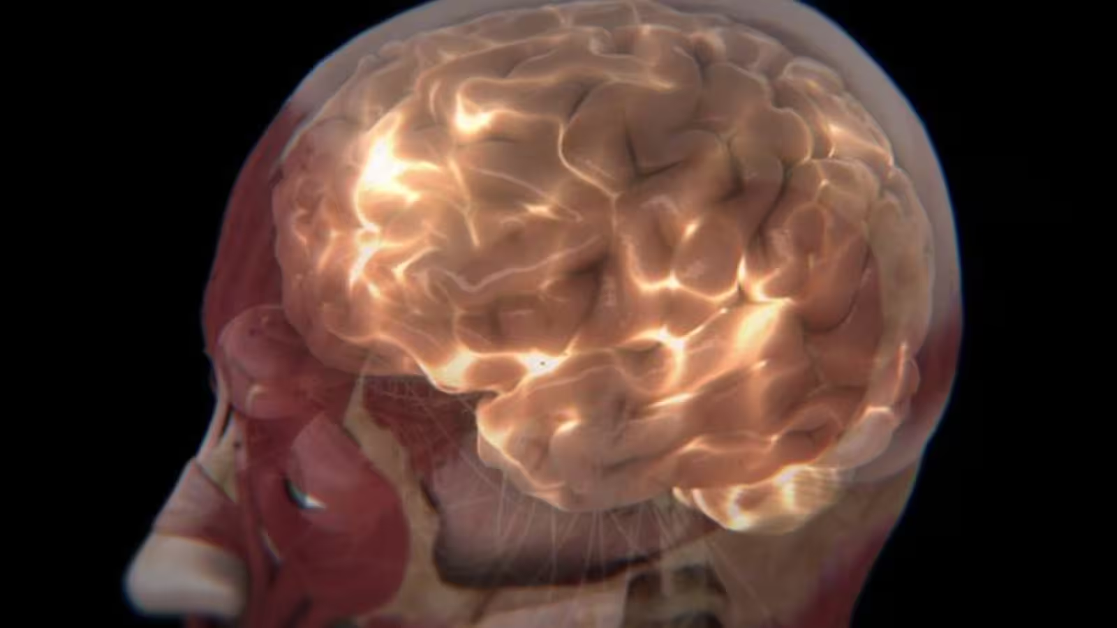
For decades, researchers have been fascinated by the phenomenon of bioluminescence in nature—a trait common among diverse organisms like fireflies, jellyfish, and certain fungi. Unlike these luminous creatures, humans are not typically associated with visible light emission. However, mounting evidence since the early 20th century suggests that human tissues do, in fact, emit an extremely faint glow. These emissions, known as biophotons or ultra-weak photon emissions (UPEs), are so subtle they evade detection by the naked eye, but modern technologies are finally enabling scientists to explore their secrets.
Biophotons fall within the near-visible to visible spectrum and are considered a byproduct of metabolic processes inside our cells. Even though all matter warmer than absolute zero emits some form of electromagnetic radiation, UPEs are distinct from ordinary thermal radiation—they represent a unique signature of biological activity.
How Is the Brain’s Light Measured? Experimental Approach and Methodology
A recent pilot study, led by biologist Dr. Hayley Casey of Algoma University in Canada, has advanced the field of biophoton research by examining UPEs from one of the human body’s most vital organs: the brain. The research team set out to detect and characterize these extremely weak photon emissions emitted by healthy human brains under different conditions, aiming to determine if these faint flashes could be linked to specific brain functions.
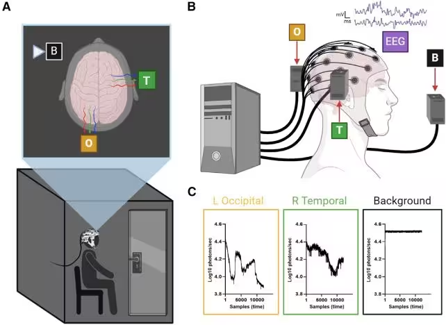
Participants in the study were placed in completely dark environments to minimize interference from external light sources. To monitor brain activity, each volunteer wore an electroencephalography (EEG) cap, which maps electrical activity across the scalp. Surrounding the participants were highly sensitive photomultiplier tubes—specialized detectors capable of capturing even the weakest traces of light.
Tests were conducted while subjects were at rest and during auditory perception tasks, ensuring all activities could occur in darkness. Across all scenarios, researchers reliably detected UPEs emanating from the head, confirming that humans continuously emit these faint photon signals. Notably, the intensity and pattern of biophoton output correlated closely with changes in EEG activity, suggesting a direct link between these emissions and the brain's functional states.
Key Discoveries: Tracking Brain Function with Ultraweak Photon Emissions
The study marks a critical step forward by demonstrating that brain-derived biophoton signals can be clearly distinguished from background noise. Importantly, for specific cognitive tasks, the UPE patterns stabilized, indicating a consistent signature associated with certain types of neural activity.
This breakthrough suggests a possible foundation for a new field: photoencephalography—a technique proposed by the researchers to assess brain health and neurological function through analysis of biophoton emissions. While EEG has long been used to track brain waves, measuring UPEs may offer a completely noninvasive window into cellular metabolism and brain dynamics without the need for electrodes or imaging tracers.
As Dr. Casey’s team reported: “We demonstrated that brain-derived UPE signals can be distinguished from background photon measures. Additionally, our results suggest that for a given task, the UPE count may reach a stable value.” This proof of concept opens up new avenues for studying the living brain.
Potential Applications and Future Directions
Researchers are now exploring how anatomical differences, health conditions, or neurological diseases might affect the intensity and distribution of brain-generated biophotons. Identifying a personal UPE “fingerprint” could enable clinicians to build baseline profiles for individuals, against which they could detect abnormal changes potentially associated with disease or brain injury.
Moreover, refining photo-detection equipment—including developing enhanced filters and signal amplifiers—will be essential for distinguishing meaningful patterns of UPEs in real-world clinical settings, both in healthy subjects and patients with neurological conditions. As the researchers note, future investigations will seek to “select filters and amplifiers to sieve and enhance UPE signal features from healthy and diseased brains,” potentially leading to powerful new diagnostic tools in neuroscience.
Conclusion
The human brain’s hidden light, undetectable to the unaided eye yet measurable with sophisticated technology, may soon transform our understanding of brain function and health. While photoencephalography remains an emerging concept, its potential to provide noninvasive, real-time insights into neurological processes is both scientifically intriguing and medically promising. As research progresses, deciphering the brain’s faint photonic language could open a new frontier in brain diagnostics—and perhaps, one day, unique light signatures will help unlock the mysteries of human cognition and consciousness.
Source: doi

Comments