
7 Minutes
Repeated soccer headers and brain health: what the new study found
Soccer, the world's most widely played sport, is under renewed scrutiny after a major research effort linked repetitive heading of the ball to measurable brain changes in adult amateur players. The largest study to date on this topic examined 352 amateur players and found that those who reported more than 1,000 headers per year exhibited microscopic alterations in a specific brain boundary layer. These structural changes were associated with modest but statistically significant declines on tests of memory and learning.
The study focused on players who had at least five years of involvement in the sport and active participation within the previous six months. Importantly, the observed effects were independent of age and sex, and they appeared in participants who had not reported concussions—suggesting that even sub-concussive impacts can be biologically consequential.
"What's important about our study is that it shows, really for the first time, that exposure to repeated head impacts causes specific changes in the brain that, in turn, impair cognitive function," said neuroscientist Michael Lipton of Columbia University, whose laboratory has led research into soccer heading for more than a decade.
Scientific background: the cortical gray matter–white matter interface (GWI)
The new results build on earlier work linking repetitive head impacts to white matter differences and to cognitive changes in contact-sport athletes. Where this study departs from much prior research is in its imaging target: the cortical gray matter–white matter interface (GWI). The GWI lies at the outermost folds of the brain—close to the cortical surface and the brain’s convoluted gyri and sulci—and has proved difficult to assess with conventional diffusion magnetic resonance imaging (dMRI).
Diffusion MRI measures the movement of water molecules in tissue and can reveal microstructural organization. Lipton’s team developed an enhanced dMRI-based method that better resolves the transition zone between gray matter (neuronal cell bodies and synapses) and white matter (axon bundles). In healthy tissue the boundary between these layers is relatively sharp; attenuation of that boundary can indicate microstructural disruption, such as altered axonal orientation, local swelling, or changes in cellular architecture.
Graduate student Joan Song, working in Lipton’s lab, described the approach: "In healthy individuals, there's a sharp transition between these tissues. Here, we studied if an attenuation of this transition may occur with minor impacts caused by heading." Using this method, the researchers identified a fuzzier boundary in players reporting high heading exposure, particularly at the anterior base of the brain.
Where the damage appears and how it may occur
The microstructural changes localized to the orbitofrontal region of the brain—just behind the eyes—an area consistent with the expected trajectory and biomechanics of a soccer header. The team hypothesizes that a contrecoup mechanism may be operating: when the skull is struck at one location, the brain can shift and collide with the opposite internal skull surface, producing focal injury. Repeated mild forces of this type, even when they do not produce a diagnosed concussion, may lead to cumulative tissue changes over time.
The orbitofrontal cortex is involved in decision-making, learning, and certain aspects of memory and emotion regulation. In this study, participants with greater heading exposure performed slightly worse on standardized memory and learning assessments. While the differences were modest on an individual level, the authors argued they are significant at the population scale, given soccer’s global participation.
The study’s authors note that damage to the GWI may have been underdetected in prior imaging studies because the cortical boundary is a technically challenging place to measure. That underdetection could help explain conflicting findings in the literature concerning whether heading causes structural brain changes in amateur players.
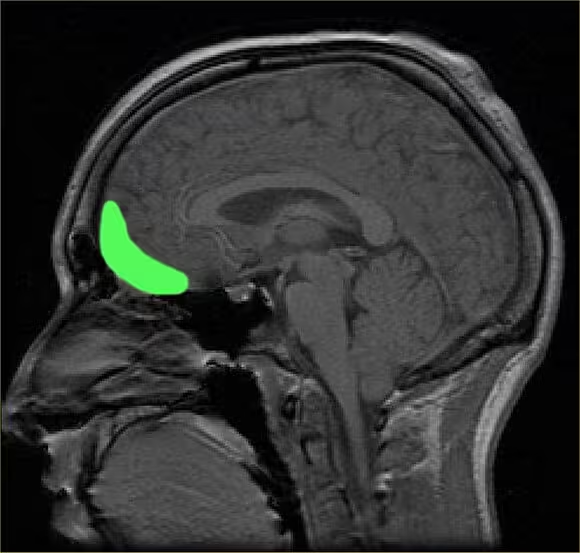
The orbitofrontal cortex, where GWI changes were observed.
Study design, methods and limitations
Key study details:
- Cohort: 352 adult amateur soccer players with at least five years of play and recent activity (within six months).
- Exposure metric: Self-reported headers per year, with a threshold of >1,000 headers used to define higher exposure.
- Imaging: Advanced diffusion MRI targeting the cortical gray–white interface to quantify microstructural integrity.
- Cognitive testing: Standardized measures of memory and learning administered to all participants.
Limitations acknowledged by the authors include reliance on self-reported heading counts, which can be imprecise; cross-sectional study design, which limits causal inference; and the need to confirm findings with longitudinal follow-up and complementary biomarkers. The team recommends prospective studies that combine objective heading measurement (for example, instrumented mouthguards or headbands), serial imaging, and long-term cognitive follow-up.
Additionally, while the GWI alterations co-occurred with cognitive differences, the effect sizes were small. That means many individuals who head frequently may experience no clinically apparent impairment in the short term, but populations with heavy cumulative exposure could be at elevated risk for subtle deficits and potentially for later neurodegenerative outcomes.
Implications for athlete safety and future research
Contact sports including American football, Australian rules football, and rugby have already faced scrutiny for the consequences of repetitive head trauma, often framed around a concussion crisis in professional ranks. Lipton’s work underscores that relatively low-level, repetitive impacts—typical in soccer heading drills and match play—may also yield measurable brain effects in non-professional athletes.
Practical implications include reconsidering youth coaching practices, high-frequency heading drills, and the development of protective strategies. Potential interventions could range from modified training to limit repetitive headers, improved technique instruction, and the adoption of monitoring technologies in high-risk contexts.
From a research perspective, the GWI now appears to be an important imaging target for future studies of sub-concussive impacts. The authors also raised the possibility that GWI alterations could relate to chronic traumatic encephalopathy (CTE) in long-term or heavy-exposure cases, though direct links to CTE require neuropathological confirmation and long-term study.
Related technologies and measurement tools
Advances in wearable sensors, head impact telemetry, and mobile biomechanics offer routes to objectively quantify heading exposure in both training and matches. Coupled with refined MRI protocols and machine-learning approaches to detect subtle microstructural changes, these technologies may enable more precise exposure–response modeling and personalized risk assessment.
Expert Insight
Dr. Elena Rossi, a fictional but realistic neuromechanics researcher and science communicator, comments: "This study advances our understanding by shifting attention to the cortical boundary, a region where repetitive, low-energy impacts can accumulate damage that traditional imaging may miss. While the average cognitive differences are small, the epidemiological signal is clear: repeated sub-concussive impacts deserve careful study and mitigation. The next step is tying objective exposure data to longitudinal brain outcomes so we can offer evidence-based guidelines to players and coaches."
What this means for players, coaches and parents
For amateur players and those involved in youth soccer, the study suggests a precautionary approach may be reasonable. Limiting high-volume heading drills, emphasizing proper technique (using the forehead rather than the hairline or side of the head), and balancing practice time with non-heading skill development can reduce cumulative exposure. Sports governing bodies and youth organizations can consider policies that restrict repetitive heading in certain age groups until more longitudinal evidence is available.
Clinicians and sports medicine teams should be aware that absence of clinically diagnosed concussions does not necessarily imply absence of impact-related brain changes. Incorporating questions about heading frequency into athlete evaluations and considering accessibility to cognitive testing or imaging for high-exposure athletes could improve early detection and management.
Conclusion
The largest study to date on amateur soccer players found that frequent heading—more than a thousand headers a year—correlates with microstructural changes at the cortical gray–white matter interface near the orbitofrontal cortex and with small but significant reductions in memory and learning performance. The research highlights a previously under-recognized imaging target (the GWI) and strengthens the case that repetitive sub-concussive impacts can accumulate into measurable brain alterations. While further longitudinal and objective-exposure studies are needed to establish causality and long-term outcomes, these findings support cautious strategies to reduce repetitive heading exposure, especially in youth and amateur settings.
Comments
No comments yet.
Leave a Comment