4 Minutes
A new electrophysiological screening method called the Fastball EEG test can detect memory-related brain changes linked to Alzheimer's disease in roughly three minutes. Rather than relying on written or verbal cognitive tasks, this test measures brain electrical responses to rapidly presented images to identify whether the brain recognises previously shown content. Early research indicates the approach may flag people with memory-specific mild cognitive impairment (MCI) who are at higher risk for developing Alzheimer’s-type dementia.
Scientific background: why early, objective tests matter
Alzheimer’s disease is a progressive neurodegenerative disorder characterized by the accumulation of amyloid and tau proteins in the brain. These proteins form plaques and tangles that disrupt neuronal communication, ultimately producing memory loss, disorientation and loss of daily function. Importantly, these pathological processes begin years before clinical symptoms become obvious, which creates an opportunity—and need—for earlier detection.
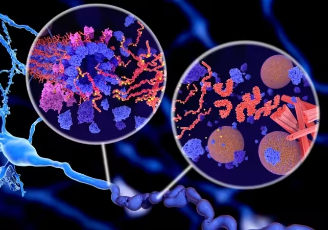
Tangles of tau proteins can trigger the death of neurons, leading to Alzheimer's disease. (selvanegra/Getty Images Pro/Canva)
Traditional cognitive screening requires active participation—remembering words, copying figures or solving problems—and is sensitive to language, education and test anxiety. Advanced biomarker tests (brain PET scans or cerebrospinal fluid analysis) provide direct evidence of amyloid or tau but are costly or invasive. The Fastball EEG test aims to bridge this gap with an inexpensive, brief and objective physiological measure of recognition memory.
How the Fastball EEG test works
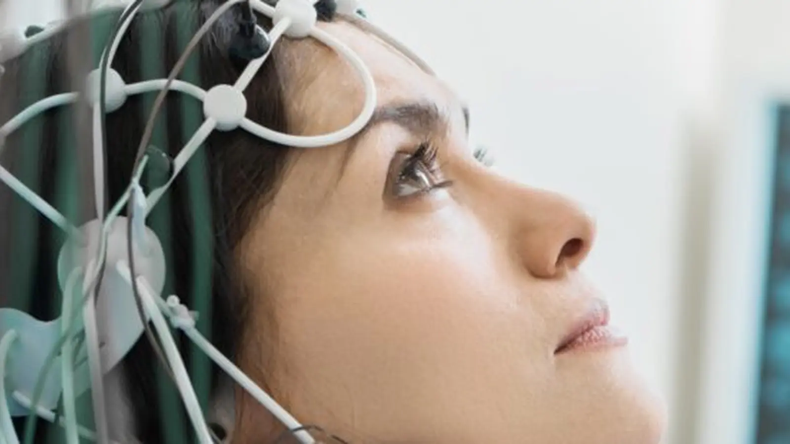
The test uses a standard EEG headset to record scalp electrical activity while images are shown on a screen at high speed (about three images per second). Before the rapid-sequence phase, participants see eight pictures that they are asked to name but not intentionally memorise. During the three-minute run, every fifth image repeats one of the eight targets among a stream of distractors. The EEG captures tiny, frequency-specific signals driven by unconscious recognition processes: strong, time-locked responses indicate intact visual recognition memory; attenuated signals suggest impairment.
Study design and key findings
Researchers evaluated 106 adults: 54 cognitively healthy controls and 52 people with mild cognitive impairment (MCI). The MCI group included both amnestic MCI (memory-predominant problems) and non-amnestic MCI (other cognitive domains affected). The Fastball EEG produced significantly weaker recognition responses in the amnestic MCI group than in healthy controls or non-amnestic MCI participants, suggesting the test can specifically detect memory-related dysfunction associated with early Alzheimer’s.
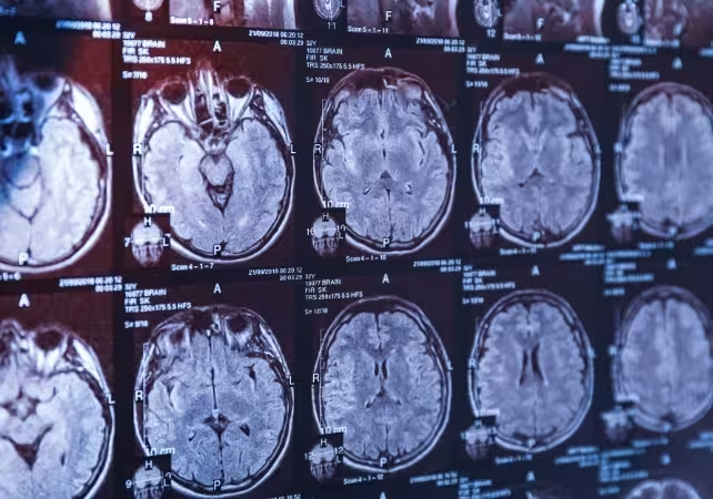
Brain scans can indicate risk of Alzheimer's, but are expensive. (pixelshot/Canva)
A subset of participants repeated testing after one year. Of 42 MCI individuals retested, eight had progressed to dementia (Alzheimer’s or vascular dementia). Standard paper-and-pencil cognitive tests did not consistently register the transition, whereas the Fastball EEG showed marginal declines in signal among those who progressed. However, the small number of converters limits conclusions about longitudinal sensitivity; larger, longer studies are needed.
Limitations and next steps
Current evidence is preliminary. The study excluded common conditions that can impair memory, such as major depression or thyroid dysfunction; therefore the Fastball EEG should not be used alone to diagnose Alzheimer’s. Important next steps include validating the test in diverse clinical populations, comparing it directly with blood biomarkers and PET imaging, and assessing performance in primary-care and home settings. If combined with emerging blood-based protein assays for amyloid and tau, the Fastball EEG could become part of a low-cost, multi-modal screening pathway to triage patients for more definitive testing.
Related technologies and implications
Blood tests that measure Alzheimer’s-related proteins are advancing rapidly and may soon allow large-scale, minimally invasive screening. The Fastball EEG offers complementary strengths: it measures brain function rather than protein burden and is portable and quick. Together, these tools could help shift dementia care from late-stage diagnosis to earlier detection and intervention, enabling lifestyle recommendations, monitoring or therapeutic trials at stages when interventions are more likely to be effective.
Expert Insight
Dr. Sarah Bennett, cognitive neurologist (fictional), comments: “Objective measures of recognition memory like Fastball EEG are attractive because they bypass language and education biases. A three-minute physiological readout that can be deployed in primary care or community clinics would greatly expand our reach. That said, sensitivity, specificity and real-world validation across common comorbidities are essential before clinical rollout.”
Conclusion
The Fastball EEG test is a promising, rapid method for detecting memory-specific brain changes linked to Alzheimer’s disease. Early research shows it can distinguish amnestic MCI from other cognitive profiles and may detect subtle declines not always captured by standard tests. While not yet a standalone diagnostic tool, the Fastball approach—especially if combined with blood biomarkers—could help identify people at risk earlier, enabling earlier monitoring, lifestyle changes and therapeutic opportunities.
Comments
No comments yet.
Leave a Comment