3 Minutes
From DIY Tool to Surgical Innovation
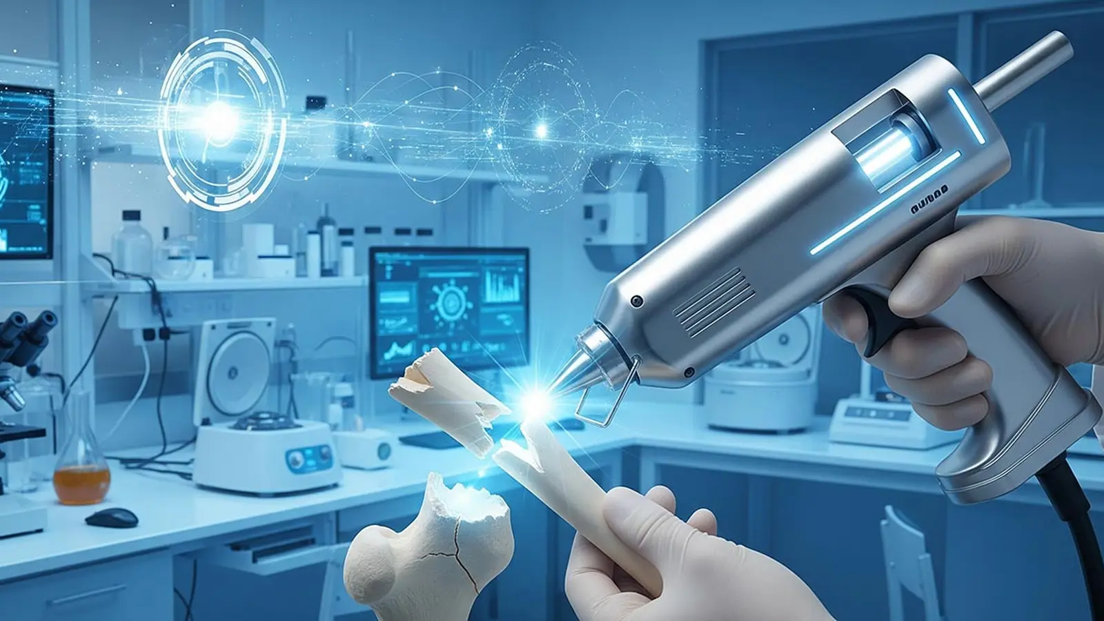
Researchers in South Korea have transformed a household glue gun into a compact, handheld device capable of producing and depositing bone graft material directly into fractures. Detailed in a recent paper in the journal Device, the modified tool enables in situ 3D printing of scaffolds onto bone defects — eliminating the need for prefabricated implants and offering a faster, more adaptable option for surgical repair.
How the Device Works
Instead of hot-melt adhesive, the team loaded the device with a tunable composite made from hydroxyapatite, a mineral closely related to natural bone, and polycaprolactone (PCL), a biocompatible thermoplastic that melts at relatively low temperatures. By altering the ratio of hydroxyapatite to PCL, surgeons can change the printed grafts' stiffness and porosity during the procedure. The compact, manually operated printer lets a clinician control printing direction, angle, and depth in real time to match irregular anatomical defects.
Product Features
- Portable, handheld in situ 3D printing mechanism
- Adjustable composite formula for on-the-fly mechanical tuning
- Biocompatible materials: hydroxyapatite and polycaprolactone
- Rapid deposition — grafting can be completed in minutes
- Designed for improved anatomical matching without preoperative modeling
Comparisons with Conventional Implants
Traditional solutions — metal plates, prefabricated polymer implants, or donor bone grafts — require preoperative imaging, modeling, custom manufacturing, or lengthy sterilization and fitting processes. In contrast, the glue-gun-style system produces scaffolds directly at the surgical site, reducing lead time and enabling a tailored fit even for complex fractures. In rabbit trials, the in situ printed grafts showed superior bone regeneration and no infection within 12 weeks compared with control animals receiving standard bone cement.
Advantages and Clinical Potential
Key advantages include reduced operative time, precise anatomical matching, and the ability to modify the scaffold's mechanical properties in real time. The research team also suggests the approach could lower postoperative complications and help curb the overuse of antibiotics by minimizing infection risks. Because the materials are biodegradable and osteoconductive, the scaffolds aim to support natural bone regrowth rather than permanently occupying space.
Use Cases and Market Relevance
Immediate applications include emergency trauma surgery, veterinary orthopedics, and settings where rapid customization is essential but advanced manufacturing infrastructure is limited. For the medical device market, the technology represents a potential shift toward mobile additive-manufacturing tools that empower surgeons with on-demand fabrication. Ongoing preclinical studies in larger animal models will determine safety, scalability, and regulatory pathways before human trials and commercial adoption.
Next Steps
The research team is expanding preclinical testing in large animals and refining material formulations and delivery ergonomics. If successful, this hybrid of low-tech hardware and advanced biomaterials could accelerate adoption of in situ printing in operating rooms, supporting faster recoveries and more personalized orthopedic care.
Comments
No comments yet.
Leave a Comment