
3 Minutes
Imagine a hidden plumbing network inside your skull, only active at night, flushing away the cellular detritus of daily thought. Sounds like science fiction. But over the past few years neuroscientists have begun to map this previously invisible system — the glymphatic pathway — and now a small Australian study suggests it might be tied to one of medicine’s most baffling illnesses.
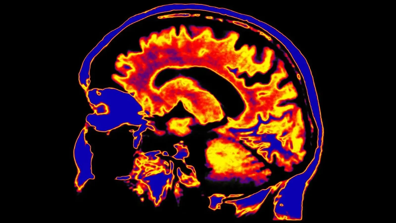
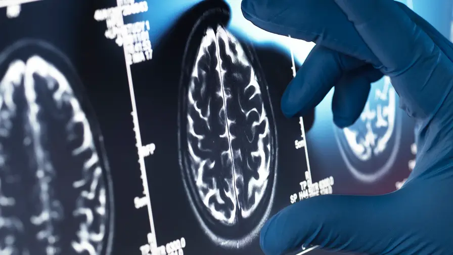
Researchers at Griffith University scanned the brains of 31 people diagnosed with myalgic encephalomyelitis/chronic fatigue syndrome (ME/CFS) and compared them with 27 healthy volunteers. They did not inject dyes or perform invasive procedures. Instead, they used a diffusion-based MRI approach that estimates how cerebrospinal fluid (CSF) moves into the tiny channels surrounding cerebral blood vessels — a proxy for glymphatic activity.

The suspected structure of the human nasopharyngeal lymphatic plexus, based on findings in mice and monkeys.
The results were striking in their specificity. Scans showed reduced markers of glymphatic function in the right hemisphere of people with ME/CFS, but not in the left. And the degree of impairment tracked with two of the illness’s hallmark complaints: troubled sleep and the so-called "brain fog" that robs patients of concentration and memory. Short. Sharp. Correlative.
Why would a clearance problem map to one side of the brain? The team is honest about that gap in understanding. Hemispheric asymmetry has popped up before in conditions such as temporal lobe epilepsy, Parkinson’s disease and ALS, yet the reason for a rightward bias remains speculative. Anatomy, vascular dynamics, sleep architecture — any (or all) of these could play a role.
What matters is the mechanism the data suggest. If the brain’s night-time cleanup crew falters, metabolic waste and inflammatory byproducts may linger in the central nervous system longer than they should. Persistent immune activation in the brain can manifest as fatigue, cognitive disruption and other neurological complaints. It’s a tidy hypothesis, and it ties together many of the biological hints that have been accumulating in ME/CFS research: immune dysregulation, altered spinal fluid markers, changes in the gut microbiome and gene expression shifts.

Diagram illustrating postmortem relocation of periarterial CSF tracers.
“This study is the first to demonstrate impaired glymphatic function in ME/CFS using MRI,” the neuroimmunologist leading the work explains, noting that the approach offers a non-invasive means to probe a system that has been notoriously hard to image in living humans. Prior direct methods required tracer injections into CSF — a technique suitable for animal studies, but impractical for routine clinical research.
Still, the team and outside experts caution against overreach. The cohort is small, the measurements indirect, and correlation does not prove causation. Does glymphatic dysfunction trigger the cascade that becomes ME/CFS? Or do systemic immune changes and disrupted sleep patterns impair the glymphatic flow secondarily? The answer is not yet clear.

Hypothesized path of CSF circulation in the human brain.
Even so, the study nudges open a promising door. If validated in larger, longitudinal studies, non-invasive imaging of glymphatic function could become a biomarker to distinguish ME/CFS from other fatigue-related disorders, guide clinicians toward targeted therapies, and focus research on restoring nighttime clearance as a therapeutic avenue.
Linking a brain-cleaning failure to chronic fatigue reframes a stubborn disease as a potentially treatable problem of clearance and inflammation.
For now the message is both cautious and energizing: a tiny, nocturnal plumbing problem may explain big, daytime suffering. The next steps are bigger studies, repeated scans over time, and experiments that test whether improving sleep or boosting clearance actually eases symptoms. Science moves slowly. But it moves.











Leave a Comment
Comments
No comments yet. Be the first.