
3 Minutes
Some spots in the brain seem to attract tumors like iron to a magnet. Strange, isn’t it? Clinicians have long noticed that certain cancers show neighborhood preferences: glioblastomas crop up in the cerebral hemispheres; medulloblastomas prefer the cerebellum in children. Pattern, not randomness. But why?
Human brains are not easy to experiment on as disease unfolds. So a team turned to an unlikely stand-in: the fruit fly. Drosophila brains follow many of the same developmental rules as ours, and researchers can push their genetics around the way you might fidget with knobs on a lab bench. That practicality revealed a surprising clue about susceptibility.
Researchers engineered fly neurons to lose the molecular marks that keep them mature and stable. The neurons reverted toward stem-like behaviors and began dividing—an established route to tumor-like masses in flies. Cells carrying the same mutation appeared across the central nervous system. But tumors did not. Only certain regions sustained growth. Others shrugged it off and returned to normal.
What made those regions different? The team tracked a protein called Chinmo, already known to steer stem-like neuronal identity. Chinmo lit up in the brain areas where tumors persisted. Where Chinmo was absent, even identical mutant cells failed to form tumors. Curious correlation. Even more curious experiments followed.

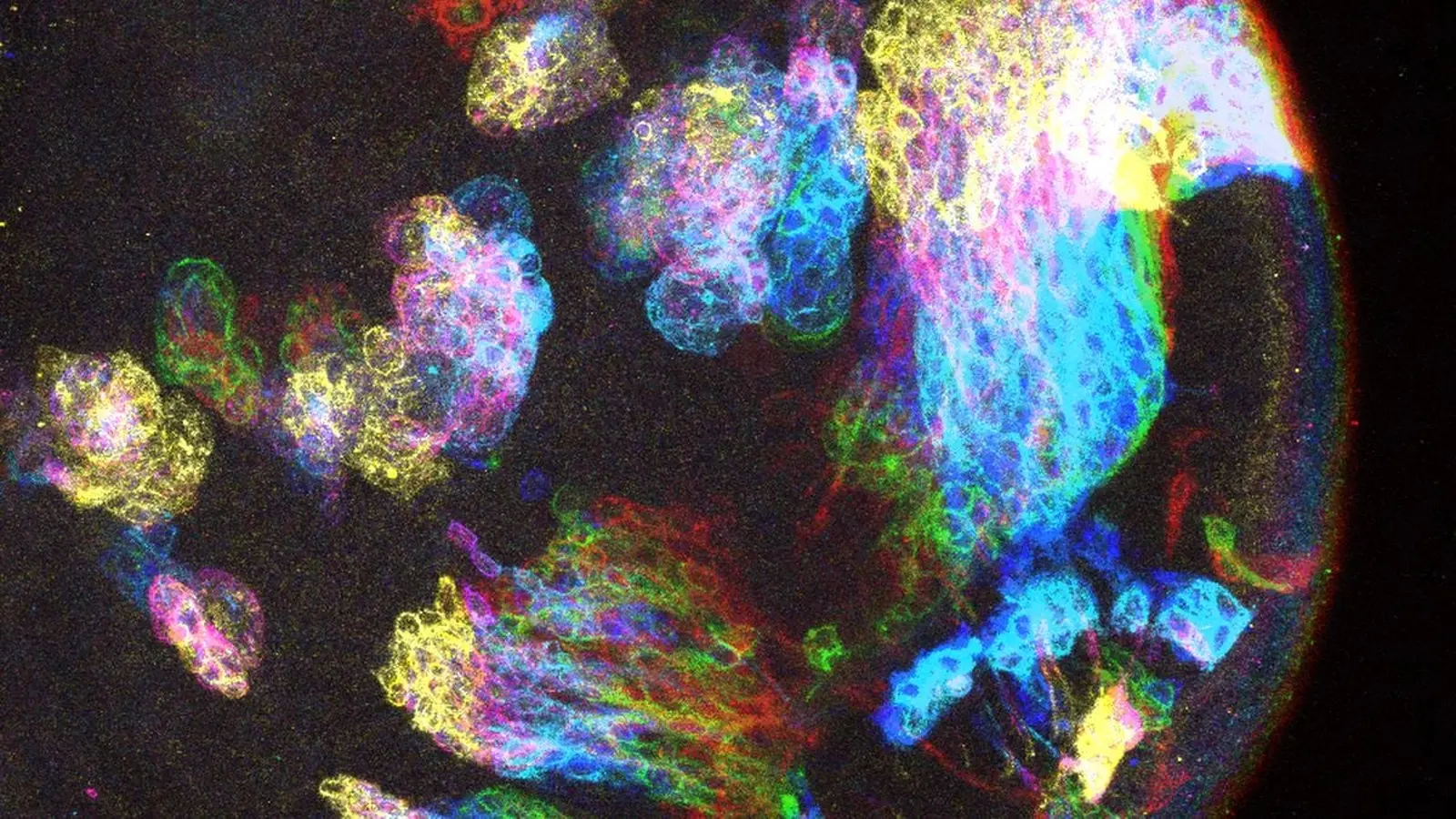
A region where abnormal cell growth (pink) coincided with Chinmo (gray)
Dial Chinmo down in tumor-prone regions and growth collapses. Crank Chinmo up in previously resistant areas and the same mutant cells begin to proliferate. Simple on-off flips, dramatic consequences. The mutation mattered, but so did the local molecular setting—the developmental state and identity signals that make a cell permissive or hostile to tumor growth.
Humans don’t carry Chinmo itself, so this is not a direct one-to-one map. Still, the result points to a broader idea: cancer risk in the brain may be shaped by region-specific proteins and programs that create windows of vulnerability. Think of it as fertile soil versus barren ground for the same mutated seed.
That shift in thinking opens new routes for therapy. Instead of targeting every mutated cell, could we change the neighborhood so those cells never take root? If human analogs of Chinmo exist—or if other local factors create permissive states—then altering those conditions could block tumors early, before they become lethal.
The study, published in Proceedings of the National Academy of Sciences, doesn’t solve brain cancer. But it gives researchers a different lens: look beyond mutations and read the map of developmental identity that may define where tumors can grow. The hunt for those maps in human tissue is already underway. What will we find when we start surveying the neighborhoods?


Leave a Comment
Comments
No comments yet. Be the first.