
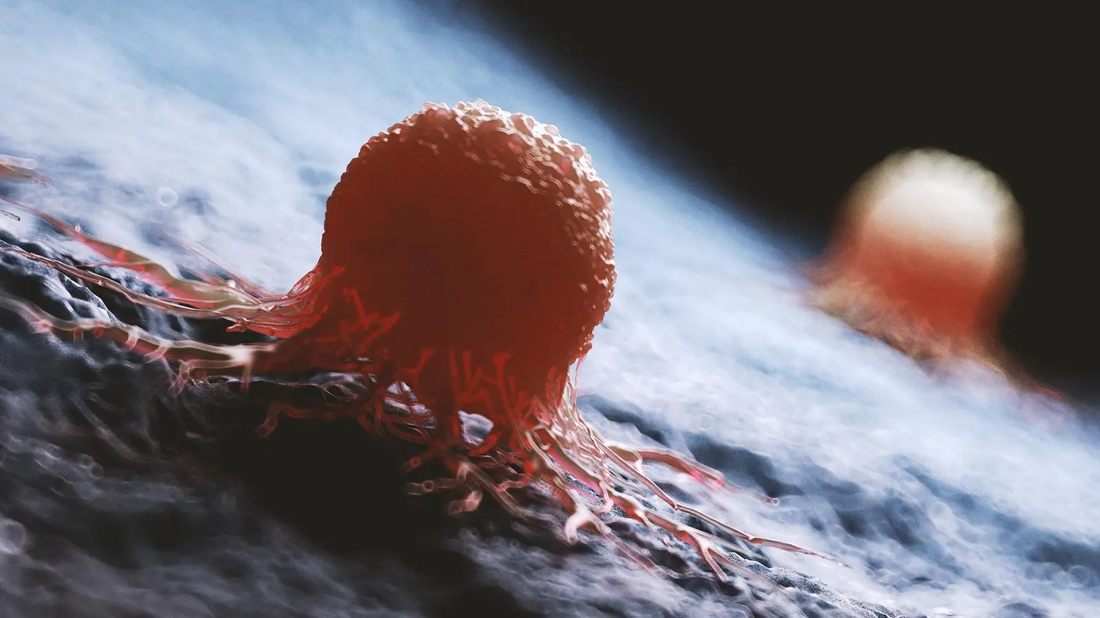
A tiny molecular change can flip a tumor from local menace to wandering killer. That flip, researchers now report, may lie in the loss of a single transcription factor: GATA6.
For patients with colorectal cancer, the real danger often begins when rogue cells seed the liver. Once that happens, therapies falter and survival rates fall. Scientists have chased obvious genetic culprits for years, looking for a mutation that explains why some tumor cells break away and thrive in a new organ. The answer from a new study is less about broken DNA and more about cells forgetting who they are.
GATA6 normally acts like a molecular librarian, keeping intestinal cells on their assigned shelf by turning certain genes on and others off. When that librarian disappears, the orderly program that defines an intestinal cell unravels. In tumor samples from patients and in mouse models, tumors that colonized the liver showed markedly lower GATA6 levels than their primary counterparts — and those low levels correlated with worse outcomes.
What follows is not mere chaos. Without GATA6, colorectal cancer cells enter a highly plastic state, a kind of cellular amnesia that lets them rearrange their identity. They flip from an LGR5-positive program to an LGR5-negative, fetal-like program — the same genetic toolkit cells use during early development or to patch wounds. That flexibility appears to help them survive detachment, circulation, immune surveillance and the foreign soil of the liver.

Immunofluorescence image of mouse colon cancer. Areas lacking GATA6 expression are LGR5-negative, indicating cells that have the potential to spread to the liver. GATA6 is shown in red, Lgr5-GFP in green and nuclei in blue.
To watch this evolution unfold, the team grew organoids from liver metastases and cycled them back into mouse colons. Tumors formed, then spread back to the liver, and by repeating that cycle the researchers could observe cells becoming progressively better at metastasizing. It was a lab recreation of Darwinian selection, but driven by changes in gene regulation rather than new mutations.
There was a surprising twist. Deleting GATA6 in models dramatically increased the frequency and size of liver metastases while leaving primary tumor growth largely unchanged. In other words, a tumor can look unchanged at the origin yet harbor cells primed for distant spread. That helps explain why tumor size and growth rate alone often fail to predict metastatic risk.
The study also supplies a practical beacon. Low GATA6 could be a biomarker that flags tumors likely to produce pro-metastatic, fetal-like cells. Restoring GATA6 activity or nudging related pathways back toward a stabilized identity reduced the cells' metastatic behavior in experiments, pointing to a strategy that aims to preserve cellular identity rather than only killing dividing cells.
Translating that idea into safe treatments will be tricky. The programs cancer hijacks are also those used by healthy tissues for repair. Still, researchers are hunting for vulnerabilities unique to GATA6-deficient cells — molecular weak spots that could be targeted without sabotaging normal healing.
Future work will also probe the tumor microenvironment: how immune cells, stromal signals and the liver's own molecular geography influence whether a wandering cancer cell successfully settles. If metastasis is a crime of opportunity, the microenvironment supplies the chance.
By shifting attention from immutable DNA changes to reversible epigenetic transitions, this work reframes how we think about metastatic risk. It suggests a new class of interventions — ones that keep cells remembering what they were meant to be.
The next step is to turn that knowledge into tests and treatments that catch the switch before the cancer makes its first trip to the liver.


Leave a Comment
Comments
No comments yet. Be the first.