
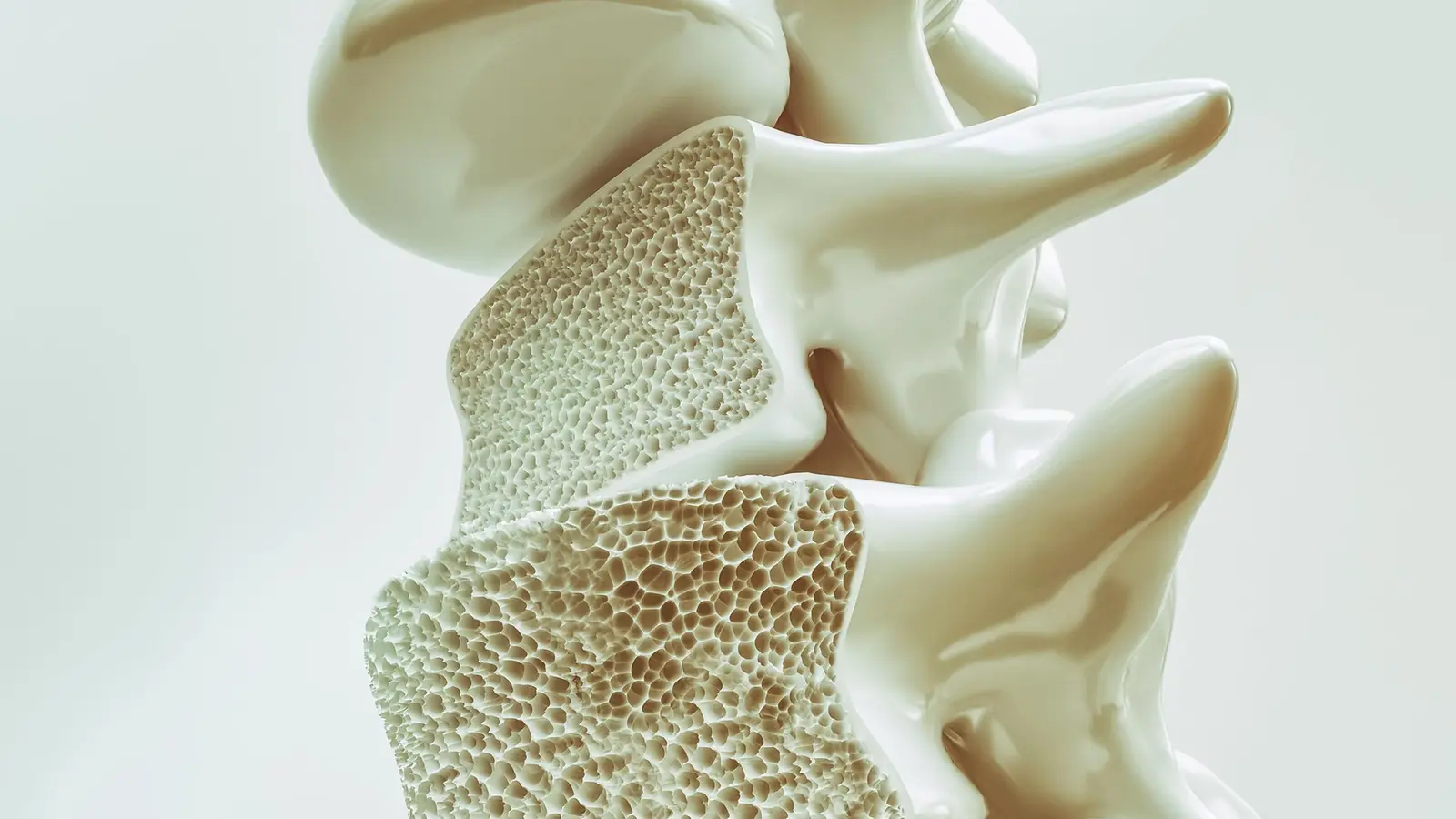
People go in for a chest CT thinking about lungs or nodules. Few expect it to whisper secrets about their brain. Yet that is precisely the surprising hint researchers are pulling from routine scans: the bones of the spine might track with how our minds age.
At Johns Hopkins, scientists dug into images from the Multi-Ethnic Study of Atherosclerosis (MESA) and found a pattern. When thoracic vertebral volumetric bone mineral density (vBMD) was lower at baseline, participants tended to show faster cognitive decline and more pronounced changes in white matter on follow-up MRI scans. The work, led by Sara Momtazmanesh and interpreted by the lab of radiologist Shadpour Demehri, used a deep-learning tool to harvest bone-density data from non-contrast chest CTs—scans already being taken for other clinical reasons.
Why look at bone when the question is brain aging? Because aging rarely hits a single organ in isolation. Bones and brains carry fingerprints of systemic processes—metabolic shifts, vascular strain, hormonal transitions—that play out across tissues. The Johns Hopkins team didn’t claim that osteoporosis causes dementia. Instead, their results point to shared aging pathways that can manifest in both skeletal fragility and declining neural integrity.
How did they measure brain change? Two imaging markers were central. White matter hyperintensities show up as bright patches on MRI and often reflect small vessel injury or tissue rarefaction. Fractional anisotropy (FA) is a diffusion MRI metric that captures microscopic organization of white matter tracts—the highways that ferry signals between brain regions. Lower baseline vBMD predicted faster accumulation of hyperintensities in the corpus callosum and a steeper decline in FA in the anterior limb of the internal capsule—areas tied to attention, working memory, and executive function.
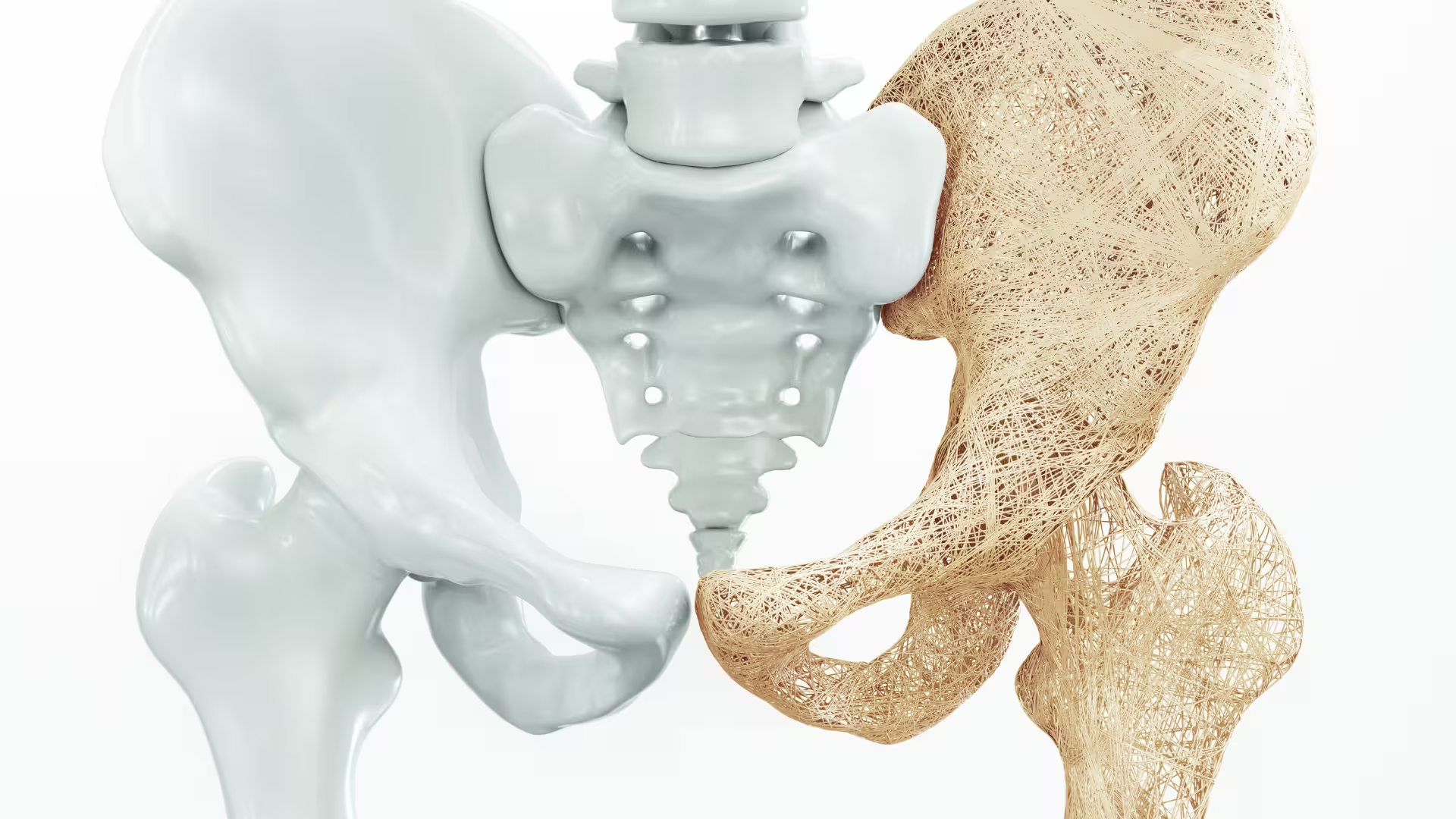
The sample that fed these findings started with more than 2,000 CTs analyzed by machine learning, narrowing to 715 people who had both the spine bone measurements and longitudinal MRI plus cognitive testing. Long-term white matter hyperintensity data were available for about 408 participants and FA for roughly 405, giving the team the ability to track structural brain change over time rather than a single snapshot.
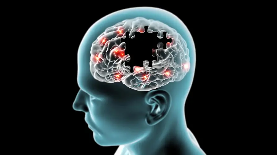
One clear result emerged: lower vertebral bone density at the outset associated with faster decline in overall cognitive performance. The imaging changes dovetailed with that clinical signal, not as a proof of cause, but as corroborating evidence that something systemic was at play.
Chest CTs performed for lung screening or other indications can offer opportunistic bone-density measurements that may flag people experiencing parallel skeletal and neural aging.
That sentence contains the practical sting. Chest CT is already routine for a host of reasons—lung-cancer screening, calcium scoring, nodule surveillance. If an algorithm can extract vBMD from those images, clinicians could gain another piece of the puzzle without additional radiation or cost. The implication is not to use bone density as a standalone dementia predictor, but to broaden risk assessment and prompt attention to shared drivers like insulin resistance, dyslipidemia, or postmenopausal changes.
There are caveats. Observational links don’t establish that bone loss drives brain injury. Confounding factors—lifestyle, vascular risk, inflammation—could underlie both processes. Still, the study offers a compelling example of how cross-organ data mining can reveal co-occurring patterns of age-related decline that were previously studied in isolation.
Beyond the specific bone–brain signal, this research showcases the rising utility of AI to synthesize diagnostic images at scale. Algorithms can now comb thoracic CTs for bone metrics, combine that with MRI-derived measures of white matter health, and relate both to cognitive tests. The result is a richer, multi-system portrait of aging.
For clinicians, researchers, and patients, the takeaway is nuanced: pay attention to the whole person. A low vBMD seen incidentally on a chest CT might prompt clinicians to consider not just fracture prevention but a broader assessment of vascular and metabolic health that also affects the brain. For researchers, the study opens pathways to probe shared biological mechanisms and test whether interventions that protect bone might also slow the course of white-matter degeneration.

We are not at the point of rewriting screening guidelines. But as imaging repositories grow and AI tools mature, the medical images already on file may become a richer source of clues about how we age—bones and brains included. Who knew a chest scan could double as a window into the mind?

Leave a Comment
Comments
No comments yet. Be the first.