

Have you ever thought of the heart as patchwork rather than a single, uniform engine? Scientists at The Jackson Laboratory did — and the picture they painted is unexpectedly intricate. By mapping thousands of individual cells from young and old mouse hearts, researchers found that the telltale signs of chronic inflammation and scarring cluster in precise anatomical neighborhoods: the aortic valves and the aortic root, not spread evenly across the muscle.
Short answer: aging in the heart is regional. Long answer: the valves and adjacent vessels—parts often dismissed as mere plumbing—appear to host concentrated molecular activity linked to inflammaging and fibrosis. Those hotspots show higher counts and altered behavior in two key support players: macrophages, the immune sentinels, and fibroblasts, the tissue’s structural repair crew.
Why does that matter? Because most cardiac aging research has zoomed in on muscle cells, especially the left ventricle, which does most of the pumping. That focus makes sense: cardiomyocytes regenerate poorly and their decline is central to heart failure. But when researchers combined single-cell sequencing with spatial genomics, the story broadened. Instead of uniform deterioration, aging revealed an uneven landscape where communication between neighboring cells shifts—and that shift is happening where the aorta meets the heart.
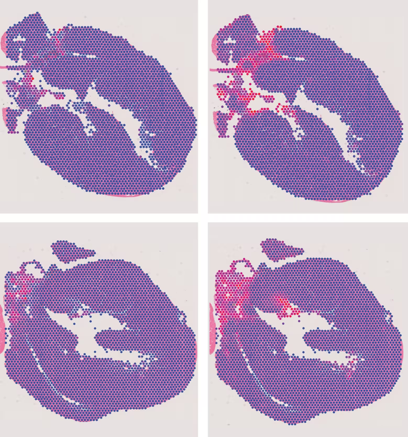
Maps of young (top) and aged (bottom) mouse hearts showing where immune cells called macrophages (left) and fibroblasts (right) are most abundant. Blue indicates fewer cells and red indicates more cells. The highest concentrations of both cell types appear around the aorta and heart valves, regions identified as hotspots of age-related change.

Macrophages in older hearts showed ramped-up inflammatory and tissue-remodeling programs. Fibroblasts, meanwhile, carried molecular signatures linked to fibrosis—excessive scarring that stiffens tissue. Two genes stood out in these hotspot zones: Ccl8, which encodes an inflammatory signal, and Cdkn1a (p21), a regulator tied to cellular senescence. But these genes didn’t act in isolation; they were part of a broader dialogue between immune and structural cells that appears to steer local inflammation and remodeling.
Here’s a curveball: some markers typically used to flag senescent cells were already present in young hearts. That surprised the team and complicates a simple interpretation where marker presence equals cell senescence. In other words, a molecular cue seen in aging tissues may play different roles depending on context and location. A six-month-old mouse heart possessing such markers is not necessarily senescent—it's a reminder that molecular signals are not always one-to-one translations of function.
Sex matters, too. The study uncovered divergent aging trajectories in male and female hearts at the cellular level. Older male fibroblasts leaned more toward fibrotic pathways, while female fibroblasts showed stronger ties to developmental and protein-production programs. These are changes in gene activity, not direct proof of functional decline, but they echo clinical patterns: men and women show different cardiovascular risk timelines and disease manifestations.
Because the mice in this study lived in tightly controlled conditions, researchers could isolate the biological effects of aging from lifestyle and environmental noise. That revealed that aging itself can ignite changes in valves and aortic tissue—regions where age-related surgical interventions, like valve replacement, are common. The findings don’t yet translate directly into therapies, but they redraw the map of cardiac aging and suggest new sites to watch.
Next steps include probing how exercise, diet, and disease alter the immune–fibroblast conversation in those hotspots. If we can separate normal, age-linked remodeling from changes that raise disease risk, interventions could be targeted more precisely—perhaps slowing valvular deterioration or taming local inflammatory circuits before they cascade into clinical problems.
Mapping where and how the heart’s cellular neighborhoods change with age opens a path toward smarter prevention—and it reminds us that the heart ages not as a single clock, but as a mosaic of clocks that tick at different speeds.

Leave a Comment
Comments
No comments yet. Be the first.