
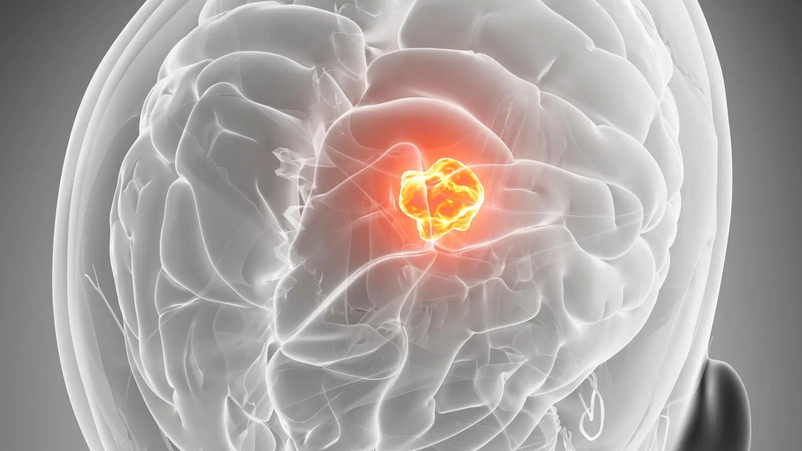
They move like restless travelers. One day a glioblastoma cell hugs a blood vessel as if following a highway. A few days later it slips into surrounding tissue, taking an entirely different route. This shape-shifting behavior is not theory. It was photographed, tracked and mapped over five days.
At Uppsala University, researchers built a workflow—called GlioTrace—that stitches together repeated microscope images and machine learning to follow thousands of individual tumor cells as they traverse living slices of mouse brain. Instead of frozen snapshots, the team captured motion: pictures taken every couple of hours, stitched into a time-lapse of cellular decisions. The result reveals a critical, and unsettling, truth about glioblastoma invasion: migration is fluid, not fixed.
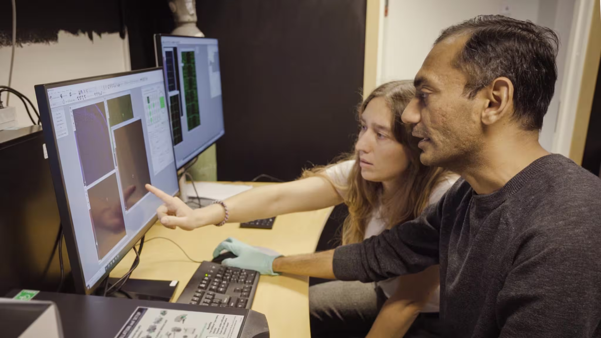
GlioTrace: Hitesh Mangukiya and Madeleine Skeppås.
Why does that matter? Because glioblastoma’s lethality stems from scattered cells that survive surgery and seed recurrence. If each cell can switch how it moves and reshape itself in days, blocking a single pathway may be like plugging one road while traffic reroutes through a dozen side streets. Some tumors favor the vascular highways; others fan out diffusely through the parenchyma. Even within a single tumor the behavior is heterogeneous—one cell’s strategy does not predict its neighbor’s.
Tracking was possible thanks to a computational pipeline that recognizes cell shapes, follows their trajectories across full brain slices, and models transitions between different movement states. The team transplanted patient-derived glioblastoma cells into mouse brains, kept thin slices alive in the lab, imaged them for up to five days, and let algorithms do the heavy lifting. That scale—thousands of tracked cells across whole slices—sets GlioTrace apart from methods that peer at only a few cells or tiny regions.

Sven Nelander, Professor at the Department of Immunology, Genetics and Pathology, Uppsala University.

Short bursts of motion. Long periods of change. Both were observed. Some cells clung to blood vessels and stayed there for long stretches; others detached and plunged into tissue, changing shape and speed within days. In some experiments, a particular fast-moving morphology sat at the center of many transitions, acting like a hub that linked different behaviors. That makes certain morphologies especially interesting—and potentially dangerous.
Treatment tests only deepened the puzzle. Two drug compounds reduced cell movement in the slices, but their effectiveness varied depending on the patient-derived tumor. And when radiation was combined with chemotherapy in some models, the population shifted toward that fast-moving cell type. Did therapy prune the slow movers and leave the fleet-footed cells behind? The study stops short of claiming causality, but it raises the possibility that common treatments change the composition of migrating cells in ways we do not yet understand.
Seeing how cells invent new routes in real time opens a path to smarter experiments and, eventually, smarter therapies.
Glioblastoma cells invading living brain tissue. Each tumor cell leaves a coloured trail showing where it moved over five days, from blue (start) to red (end). Blood vessels and immune cells (microglia and macrophages) are shown in grey. Some tumor cells migrate along the blood vessels, while others move freely through the tissue. The image was recorded with GlioTrace, a new method that uses artificial intelligence to follow thousands of individual tumor cells in living brain tissue and measure how they switch between different ways of invading.
GlioTrace’s software, developed in collaboration with teams at Chalmers University of Technology and the University of Gothenburg, is available to other labs. That means researchers worldwide can replicate the approach, compare tumors from different patients, and test how candidate drugs influence migration networks rather than single behaviors. For now, the technique is a research tool—not a clinical diagnostic—but its release accelerates investigation into how cells interact with blood vessels, immune cells and the brain’s structural matrix.
There are still big unknowns. Are the fast-moving cells more treatment-resistant? Do they seed recurrence more effectively than their slower peers? Answering those questions will require follow-up studies that connect migration patterns to molecular profiles and long-term outcomes. For now, GlioTrace reframes the problem: glioblastoma invasion is not a single tactic to outwit but a shifting repertoire to understand.
If tumor cells are improvisers, our experiments must become choreography—tracking movement, predicting switches, and testing interventions that anticipate not just where cells go, but how they change along the way.

Leave a Comment
Comments
No comments yet. Be the first.