
Some pulses land like a bell; others barely whisper. That inconsistency has bedeviled brain stimulation for years — the same electrical or magnetic tweak to the cortex can spark a robust response one moment and almost nothing the next. New work using detailed recordings curated by EBRAINS suggests we may have been looking in the wrong place: it’s not just where you stimulate the brain, but what the brain is doing at the split second before the pulse arrives.
Researchers mined an EBRAINS-hosted dataset — simultaneous intracranial SEEG and high-density scalp EEG from 36 people undergoing medically necessary mapping for epilepsy — and treated the data as a kind of cinematic record of brain dynamics. Across roughly 320 sessions and more than 10,000 single-pulse electrical stimulations, the team computed 125 pre-stimulus measures spanning network synchronization, functional connectivity, and signal complexity to ask a simple but bold question: which aspects of the brain’s immediate state predict how it will react?
The short answer: the whole-brain context matters. Local recordings alone were blunt predictors. When the scientists widened their lens to include distributed activity patterns, predictive power rose substantially. Predictions were strongest for stimulations aimed at sensorimotor and visual networks, suggesting some circuits are more tightly gated by their ongoing rhythms than others.
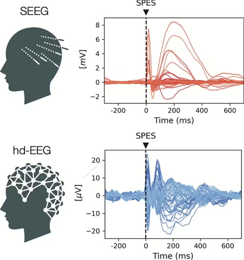
Responses to single-pulse electrical stimulation (SPES) in one study participant, recorded via SEEG) hdEEG. Brain stimulation is used in therapy and research and applies electrical or magnetic fields to modulate neural activity.
Perhaps the most practical finding is that trimming trials to those with particular pre-stimulus signatures reduced response variability by up to 24 percent. That’s not an academic nicety; it’s a tangible reduction in noise that could make neuromodulation effects more consistent across trials and patients. Importantly, the informative patterns showed up in both intracranial SEEG and noninvasive scalp EEG, hinting that clinical applications need not rely solely on invasive monitoring.
Why does this matter? Neuromodulation is increasingly used to probe circuits and treat conditions such as depression, Parkinson’s disease, and epilepsy. Yet its clinical adoption is limited by unpredictability: two identical settings can produce very different outcomes. If clinicians could monitor a patient’s brain state in real time and time stimulation to moments when the network is primed to respond, the therapy could shift from probabilistic to calibrated.
Beyond the clinical angle, the study underscores the value of open, well-documented datasets. EBRAINS enabled a reproducible, large-scale analysis that would be hard to assemble in a single center. The methods here point toward adaptive stimulation paradigms that read the brain’s immediate signatures and decide — in milliseconds — whether to deliver a pulse.
We are still some way from a routinely used, brain-state–aware device in the clinic. But the idea is simple and enticing: listen first, nudge second. If we learn to tune stimulation to the brain’s brief windows of receptivity, neuromodulation could become not just more reliable, but more precise and personalized than ever before.

Leave a Comment
Comments
No comments yet. Be the first.