
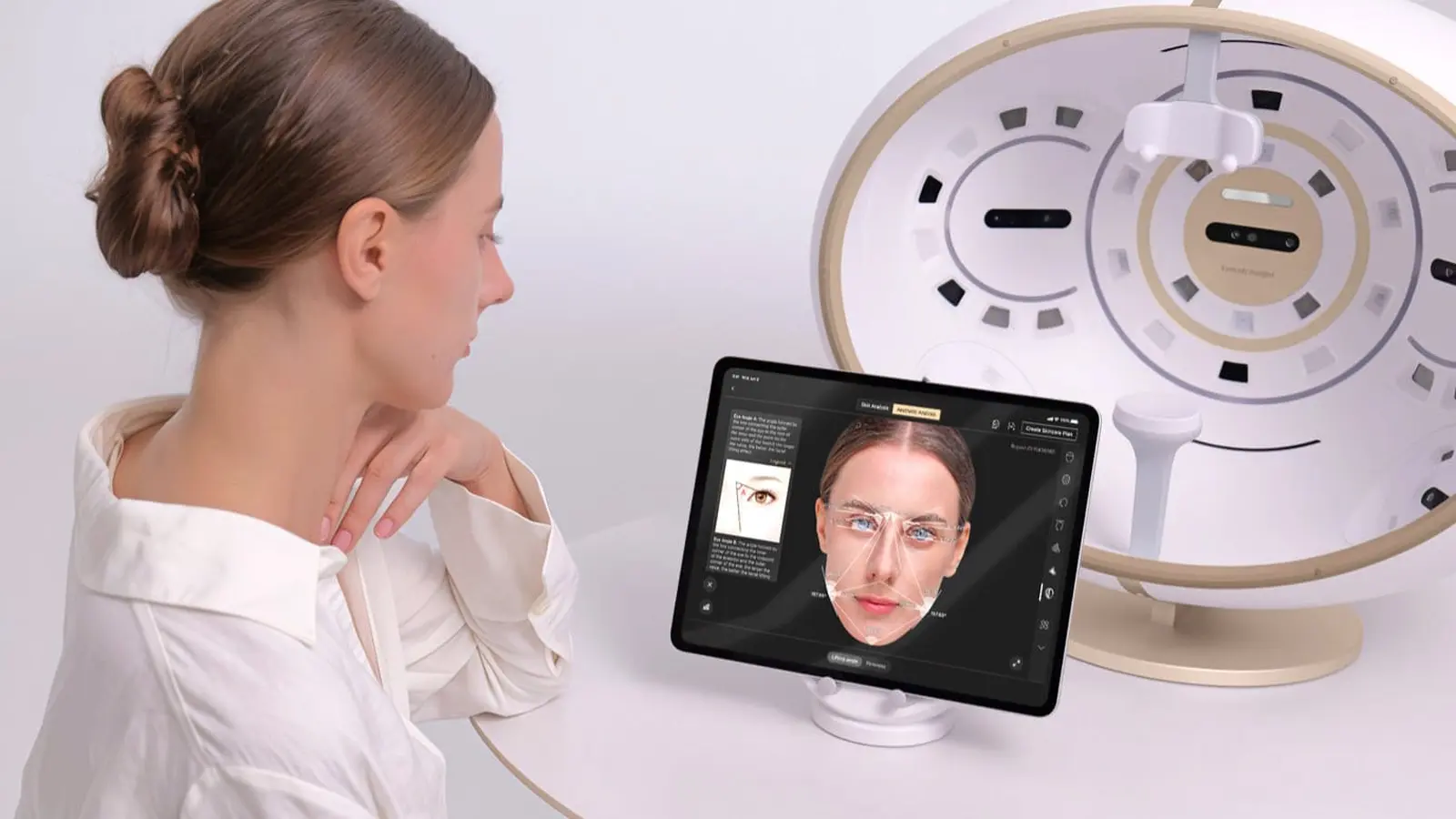
Imagine a device that reads the skin like an X-ray reads bones — except its resolution is measured in micrometers and it works in real time. Researchers at Friedrich-Alexander-Universität Erlangen-Nürnberg are testing precisely that: a combination of ultra-high-resolution imaging and artificial intelligence that flags tiny basal cell carcinomas long before they become obvious to the naked eye.
The technique at the center of the study is called line-field confocal optical coherence tomography, or LC-OCT. Think of it as two complementary microscopes in one: optical coherence tomography maps how deep a lesion goes, while confocal microscopy teases out cellular detail. The result is vertical, horizontal and 3-D images of skin structures at a scale of thousandths of a millimeter — fine enough to see details a human hair would dwarf.
AI sits alongside the scanner, not in place of the clinician. As images are captured, the software overlays a color-coded probability map of where basal cell carcinoma might lurk. It’s a prompt. A nudge. The doctor still makes the diagnosis, but the algorithm helps point the eye where it matters most.
In a feasibility study of 150 people considered at higher risk, a systematic facial screening protocol dubbed SUBSCAN turned up 14 histologically confirmed, subclinical tumors — roughly 9.3% of participants. Eighteen lesions were flagged by the AI-assisted scans; biopsies later confirmed 15 of those as basal cell carcinoma. Two patients declined biopsy, and one lesion was an actinic keratosis. That yields a positive predictive value of about 83.3% for lesions the system labeled as cancerous and were subsequently sampled. What the study did not establish, however, is how many cancers the method might miss — its sensitivity remains an open question.
Basal cell carcinoma develops from basal cells in the outermost layer of the skin and is strongly linked to cumulative ultraviolet exposure.
Most of the detected growths were superficial basal cell carcinomas; investigators also found nodular tumors and a single infiltrative case. That pattern matters because superficial lesions are the ones most likely to be treated noninvasively — with creams or topical therapies — rather than excised. Early identification can therefore change the clinical pathway, reducing scarring and the need for surgery, particularly on delicate facial structures.
LC-OCT’s ability to render cellular detail in real time is what makes this approach promising. Where a standard skin exam depends on visual clues and a clinician’s experience, LC-OCT reveals architecture beneath the surface. Add an AI that highlights suspicious areas as imaging proceeds, and you get a targeted, faster triage of sites worth biopsy or monitoring.
But practicality remains a hurdle. SUBSCAN requires systematic, careful imaging of facial skin that appears normal, and at present the process is time-consuming. The method shows promise as a screening adjunct for people at high risk, yet it is not ready for broad, routine use. Larger studies are needed to define how often it misses cancers, how it performs across different skin types, and whether workflows can be streamlined so that clinics can adopt it without bottlenecks.
There are larger questions, too. Could bringing subclinical cancers into view reshape treatment algorithms so topical therapy replaces surgery more often? Will this kind of targeted imaging reduce long-term morbidity on cosmetically sensitive areas like the nose and around the eyes? Technology can reveal what was invisible; how medicine uses that vision will be the next decision to make.
If SUBSCAN and AI-assisted LC-OCT clear further clinical hurdles, the future of skin cancer screening might feel less like guesswork and more like guided inspection — a precise, early look beneath the surface that gives patients simpler options and clinicians better information.

Leave a Comment
Comments
No comments yet. Be the first.