
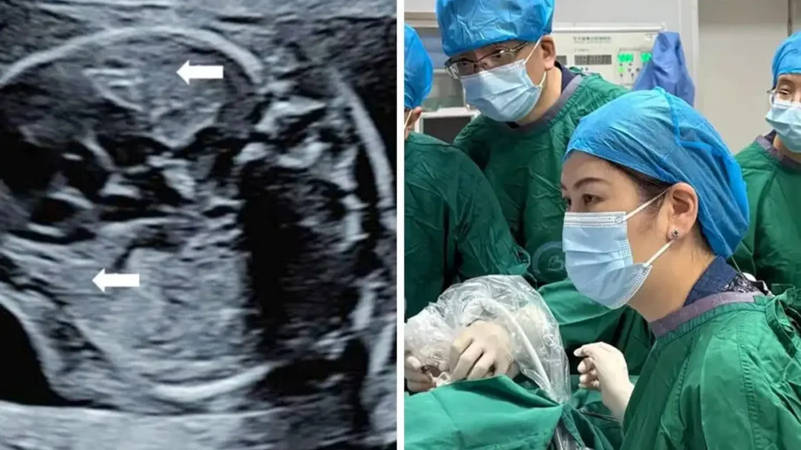
They threaded a hair-thin probe through the mother's abdomen and, under live ultrasound, aimed at a living tumor the size of a small plum lodged in the fetus's chest. The scene sounds like science fiction, but it is a painstaking, real procedure carried out by clinicians aiming to rescue a baby before it ever took a first breath.
A multidisciplinary team from Bao'an Shenzhen Women and Maternity Hospital and The Chinese University of Hong Kong reported the intervention in Ultrasound in Obstetrics & Gynecology. The patient was a 40-year-old woman at 22 weeks' gestation. Prenatal scans had revealed a rapidly enlarging congenital pulmonary airway malformation (CPAM) in the right hemithorax—microcystic in appearance, roughly 5 cm across, compressing the heart and lungs and accompanied by ascites. Left unchecked, this constellation often progresses to fetal heart failure and hydrops fetalis, a life-threatening fluid accumulation.
Typical fetal therapies—such as draining a dominant cyst or occluding an arterial feeder—work when a tumor presents a clear target or a feeding vessel. But this lesion was different: a dense cluster of tiny cysts with no obvious central blood supply. Conventional strategies offered little hope. So the team took a different tack: direct thermal ablation from inside the womb.
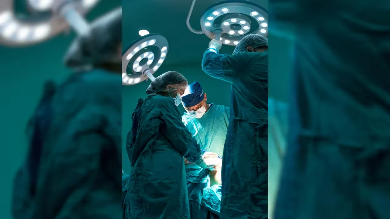
Under continuous, high-resolution ultrasound guidance, surgeons advanced an ultra-fine ablation needle through the maternal abdominal wall, the uterine wall, and into the fetal chest to reach the tumor core. The device used microwave energy to agitate water molecules in the lesion, generating intense, localized heat that coagulated and destroyed the abnormal lung tissue. The active heating phase lasted only about two minutes, and temperatures were tracked in real time on monitors to protect surrounding fetal organs.
Within three days the mass had collapsed and the excess fluids resolved. The turnaround was dramatic. The fetus stabilized, the pressure on the heart eased, and the progression toward hydrops stopped.
The pregnancy continued to term. At 37 weeks the child was delivered by cesarean, weighing 2.9 kilograms and scoring a perfect 10 on the Apgar test. At a four-month follow-up there were no signs of recurrence and no additional surgery had been required—an outcome that, if sustained, marks a major step forward for in-utero care of selected pulmonary malformations.

Why does this case matter? Aside from the immediate human story—a life redirected before it began—it demonstrates that focused microwave ablation can be safe and effective for certain microcystic fetal lung lesions that elude standard approaches. It does not replace existing techniques, but it adds a valuable option when anatomy rules out shunts or vascular occlusion.
There are caveats. Single-case reports require cautious interpretation. Long-term respiratory outcomes, risks of thermal injury, and reproducibility across centers must be studied. Still, the procedure opens a doorway. Clinicians and researchers will be watching closely to see whether this method can be refined, scaled, and integrated into fetal therapy programs worldwide.
Could this be the start of a new chapter in prenatal surgery—and a new lease on life for babies with otherwise fatal lung lesions?

Leave a Comment
Comments
No comments yet. Be the first.