
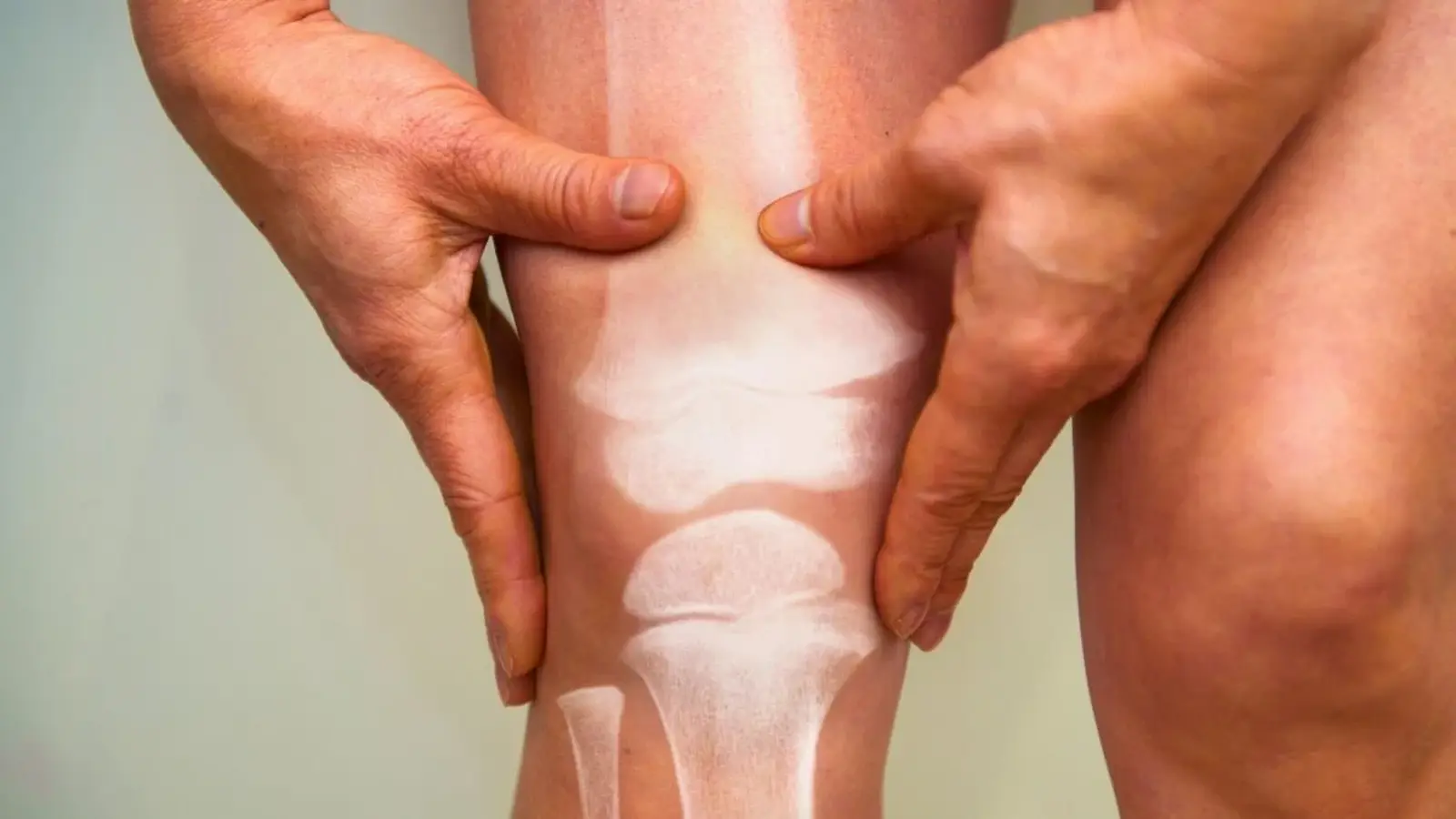
What if a routine scan for your lungs could whisper a warning about your brain years down the line? That provocative possibility is the headline from a new Johns Hopkins-led analysis that repurposed chest CT images to read bone health—and found ties between weaker vertebral bone density and signs of faster brain aging.
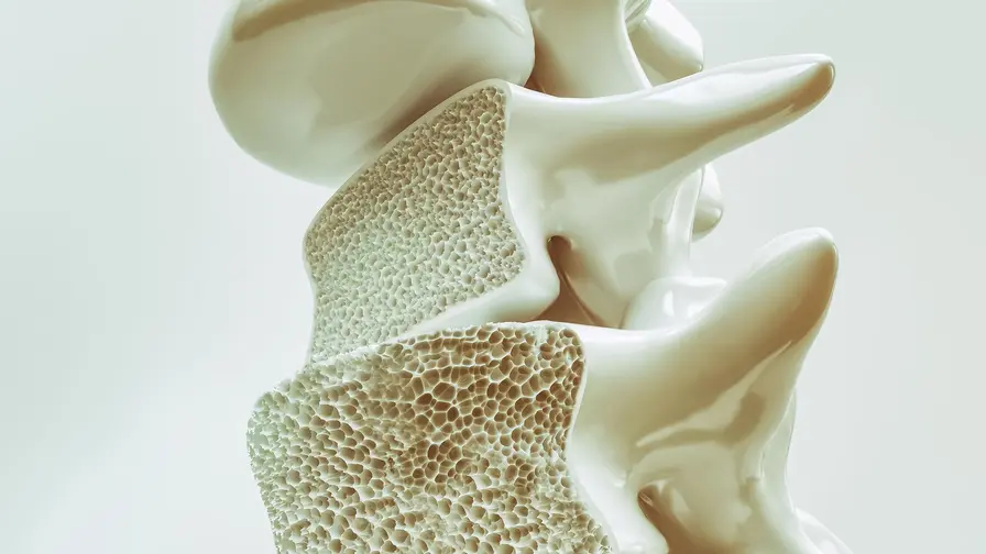
Researchers sifted through existing data from 715 participants enrolled in a long-term health study. Those people had undergone chest CTs and cognitive testing at baseline, then returned years later for follow-up thinking and memory exams and brain imaging. Instead of ordering fresh tests, the team let a custom-trained AI comb the archived chest scans for bone mineral density (BMD) in the thoracic spine. Less mineral content—calcium and friends—means weaker, more fracture-prone bone. Simple as that. Or so it might seem.
The surprising pattern was not bone fractures but brain changes. Lower BMD at baseline was associated with two markers of brain wear: growing white matter hyperintensities, which appear as bright lesions on MRI and flag damaged tissue, and reduced fractional anisotropy in white matter tracts, a diffusion MRI metric that signals diminished structural integrity. In plain language: the participants with weaker vertebral bones tended to show imaging signs that neuroscientists link to faster brain aging—and they also scored worse on cognitive tests over time.
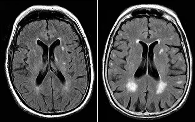
White matter hyperintensities show up as bright white areas on scans, and indicate brain tissue damage.
"This study is the first longitudinal secondary analysis linking baseline vertebral bone mineral density to changes in white matter structure, white matter hyperintensity progression and cognition," says radiologist Shadpour Demehri of Johns Hopkins. The paper, published in Radiology in 2026, leans on the combined power of imaging and clinical follow-up to make a case that a snapshot of bone strength may map onto both functional and structural measures of age-related brain decline.
Does that mean weak bones cause the brain to falter? Not remotely. The team is careful about that. "This study is not about cause and effect, but rather the observation of a metabolic syndrome that may cause both bone and brain degeneration," Demehri adds. In other words, the two may be co-travelers on the same biological road—driven by shared forces such as insulin resistance, abnormal lipids, and the hormonal shifts of menopause—rather than a direct bone-to-brain line of fire.
Still, the correlations point to specific brain regions. The white matter lesions and reduced fractional anisotropy clustered in areas tied to executive function, attention, and working memory—networks that let you plan, hold information in mind, and focus. It's those everyday mental skills that slipped more quickly among people with lower vertebral BMD.
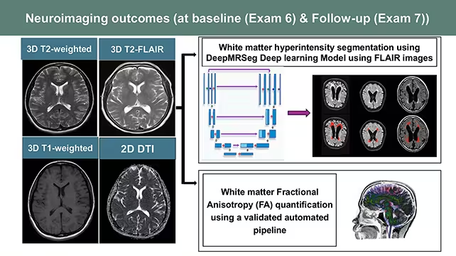
The researchers looked for two different markers of brain wear and tear.
Beyond the clinical associations, the study also showcases a practical idea: latent data is a gold mine. "Diagnostic images contain an immense amount of data that AI can now synthesize at scale across multiple organ systems," Demehri notes. With modest extra training, chest CTs acquired for lung cancer screening, cardiac calcium scoring, or nodule follow-up could yield opportunistic bone-density measurements from the thoracic spine. Sara Momtazmanesh, a coauthor and fellow radiologist at Johns Hopkins, highlights this upside. These scans, she argues, could provide an opportunistic measurement of bone density that correlates with cognitive trajectories.
That promise comes with caveats. The cohort numbers—715 people—are solid for an imaging study but not definitive for public-health screening. Follow-up covered several years, which strengthens the temporal link, yet longer observation is needed to know whether the faster brain aging flagged here translates into clinically diagnosed conditions such as dementia. And predictive screening would demand far more validation across diverse populations and scanner types before medical practice shifts.
There is, too, a larger scientific opportunity. If different age-related maladies share metabolic roots, then treating the upstream driver might buy benefits across organ systems. Think of it as addressing a river’s headwaters rather than mopping up downstream floods. But first, researchers must untangle which metabolic pathways truly unite bone loss and brain degeneration—and whether interventions that shore up bone can also slow cognitive decline.
For now, the take-home is part cautionary tale, part blueprint. Existing clinical imaging stores are not inert archives; they are living datasets waiting for smarter analysis. With AI, we can repurpose a single scan to reveal clues about multiple organs. The next step is rigorous testing—longer studies, broader cohorts, and careful work to separate association from causation—so that those clues can be turned into reliable tools for prevention and care.
After all, medicine advances when we start asking new questions of old records—and when a scan meant for one problem quietly points us toward another.

Leave a Comment
Comments (1)
Wow didnt expect chest CTs could hint at brain aging... kinda spooky 😮, hope this holds up, and more studies pls!