
4 Minutes
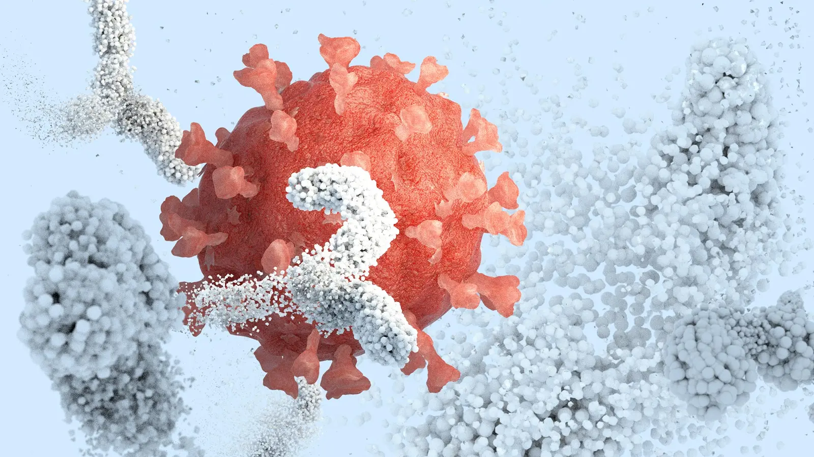
They were missing the stage. For years, scientists studied viral surface proteins like actors photographed offstage—costume intact but without the set that shapes their posture and moves. The result: snapshots that missed how antibodies actually approach, grip, and disable a virus in the field.
Now a team led by researchers at Scripps Research, in collaboration with IAVI and others, has rebuilt part of that set. By embedding viral proteins into tiny lipid discs—nanodiscs that mimic a virus’ membrane—they can watch antibodies interact with these proteins in a setting that more closely resembles a real virus. The images and functional tests that follow make some longstanding mysteries suddenly visible.
Why does this matter? Because many vaccine efforts rely on lab-made protein fragments that leave out the membrane-anchoring portions. Those omitted segments are not mere tailpieces; they influence how the entire protein folds, how it sits on the viral surface, and how nearby antibodies can reach it. Without that membrane context, researchers can miss key contact points and misjudge which antibody responses are protective.
Nanodiscs solve that problem in a pragmatic, scalable way. Imagine cutting a small, stable patch of a cell membrane and gluing a viral protein into it so the protein behaves like it would on an actual virion. Lipids provide the scaffolding; the nanodisc keeps the protein oriented and constrained. The platform preserves structure, supports common laboratory assays, and adapts to high-resolution imaging tools. Shorter experiments. Cleaner data. Better comparisons between vaccine candidates.
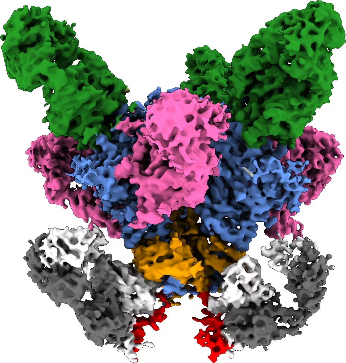
A viral surface protein (blue and orange) is shown bound to multiple antibodies (pink, green and gray/white), with a region near the membrane (red).
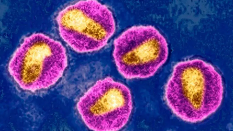
The team tested the approach on tough targets: HIV and Ebola. Both viruses present surface proteins that are notoriously tricky for the immune system to spot and neutralize. With HIV, researchers focused on a conserved region near the membrane base—an area that some broadly neutralizing antibodies target. When that region was presented within a nanodisc, cryo-EM and binding assays revealed interactions that simply don’t appear when the protein is stripped free of its membrane anchor.
Those newly visible contacts explain, in structural terms, why certain antibodies can block a wide swath of viral variants. Some antibodies work by prying apart protein assemblies or by inserting themselves at the membrane interface to destabilize the machinery the virus needs to fuse with cells. Seeing that choreography up close gives vaccine designers a clearer idea of what to mimic when they want to coax the immune system into making similarly effective antibodies.
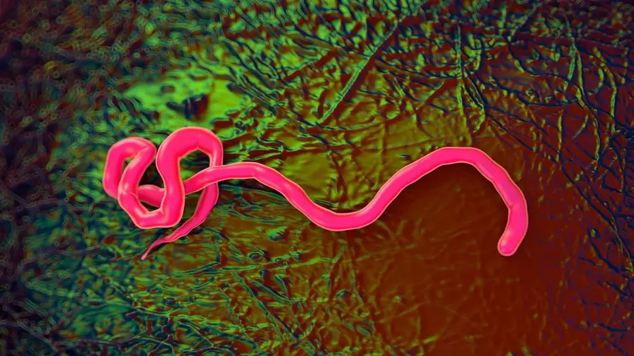
And the approach isn’t limited to HIV. The same membrane-mimicking strategy worked with Ebola surface proteins in the lab, suggesting broad applicability to other membrane-anchored viral antigens, from influenza hemagglutinin to SARS-CoV-2 spike regions that lie near the viral envelope.
There’s also a practical upside for vaccine discovery workflows. Nanodiscs can act as molecular bait, capturing B cells and antibodies that recognize the membrane-contextualized antigen. Experiments that once took a month can be compressed into a week, accelerating the pipeline from candidate design to comparative evaluation. For teams racing to test many constructs, that kind of speed matters.
The platform itself is not a vaccine; it is a more faithful lens through which to design one.
Building this system involved bringing together existing pieces—lipid chemistry, protein engineering, and imaging—into a reproducible, scalable package. That integration is what transforms an intriguing lab trick into a usable research tool. The payoff is both conceptual and actionable: unexpected antibody footprints become visible, and those footprints can be used as blueprints for next-generation immunogens.
Science often advances when we stop simplifying reality for convenience and start replicating its messy details. Nanodiscs do just that for membrane-bound viral proteins. They restore a missing context and, in doing so, reveal why some antibodies succeed where others fail. For vaccine developers chasing the broad, durable protection seen only rarely in nature, that extra context could be the difference between guesswork and design.
Think of it as giving immunologists a more honest mirror—one that reflects how a virus really looks to the immune system, not a staged approximation. The next step will be to translate those structural revelations into immunogens that reliably elicit the protective antibodies now visible in unprecedented detail.

Leave a Comment
Comments (2)
Is this even real? Seems clever but also could be an in vitro trick, like will it mimic real virions or just a neat lab toy... if that's real then huge, but skeptical
wow didnt expect the membrane to change everything. seeing antibodies wedge into a tiny lipid patch gave me chills... if this scales, big deal