

5 Minutes
Imagine a single molecular switch flicking on in the wrong place and setting off a neighborhood-wide blackout. That image helps explain a bold new idea about how Alzheimer’s may begin not as plaque alone, but as a local miswiring inside particular neurons that then drags the surrounding tissue into decline.
The hallmark images of Alzheimer’s — amyloid plaques and tau tangles — tell only part of the story. Long before memory problems dominate clinic notes, neurons can start misfiring, synapses vanish, and glial cells turn defensive. How these disparate events link together has been a puzzle. A team led by Won‑Suk Chung at the Institute for Basic Science thinks they’ve found a molecular fulcrum: the receptor ERBB4, appearing where it shouldn’t.
ERBB4 normally lives in inhibitory neurons, where it helps apply the brakes on brain activity. But using single‑nucleus RNA sequencing in mouse models, the researchers discovered a small, early‑emerging group of excitatory neurons that oddly express ERBB4. The team named them Early Responsive Excitatory Neurons — ERENs. What looked like a curious marker turned into a suspect driver when manipulating ERBB4 changed the disease course in striking ways.
Glial cells — astrocytes and microglia — are the brain’s maintenance crew. They prune synapses, clear debris, and help sculpt circuits during learning. In the Alzheimer's models the IBS team studied, these glia began selectively removing excitatory synapses while sparing or even preserving inhibitory ones, tilting the balance toward runaway activity. That selective pruning amplified circuit instability rather than calming it.
Was the cleanup crew to blame, or were they reacting to a cry for help? The evidence points to the latter. When neurons were experimentally driven to fire more, glia increased their synapse engulfment. Quiet the neurons, and the pruning subsided. The pattern suggested that damaged or hyperactive neurons were signaling distress, and glia were responding — sometimes destructively.
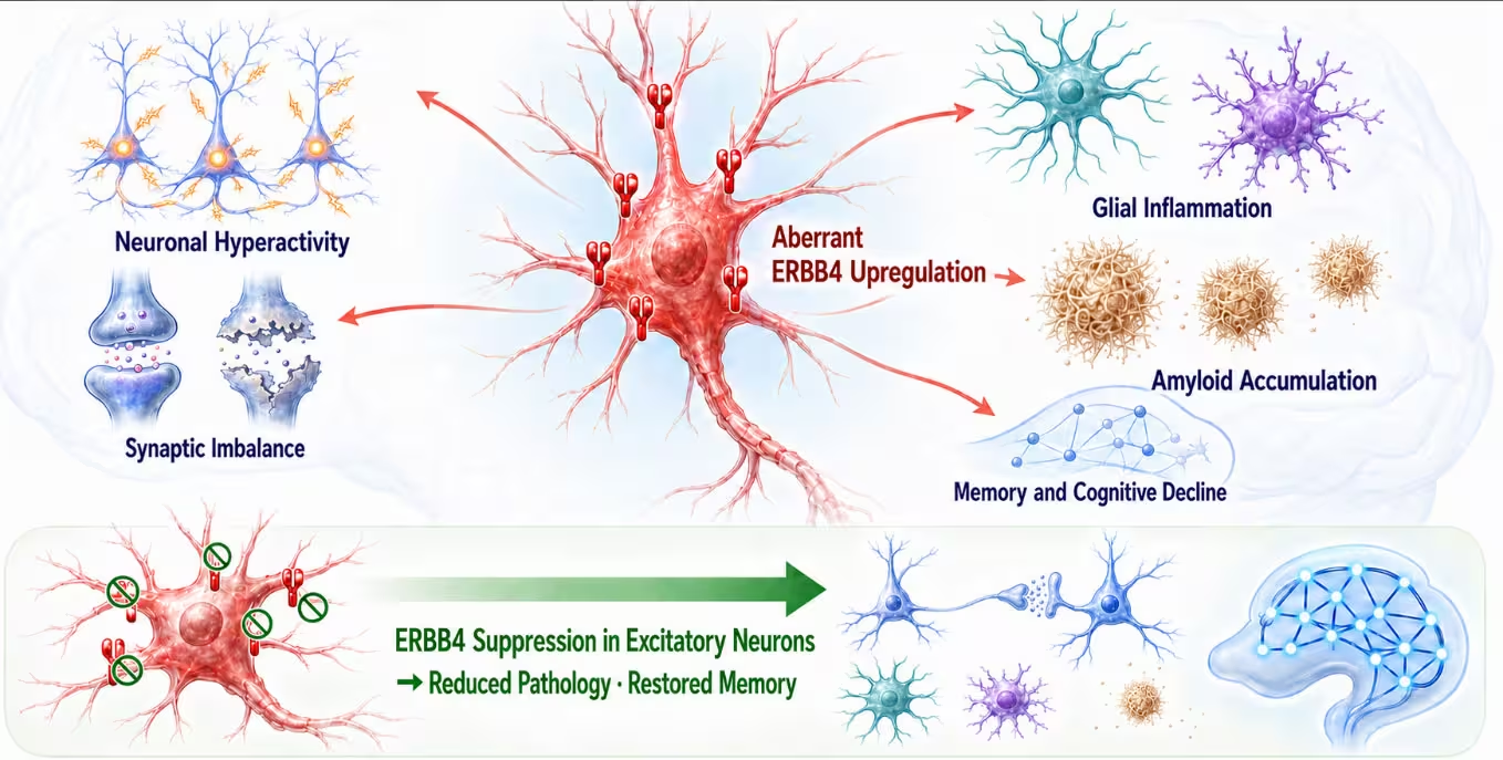
Schematic illustration of the proposed disease mechanism whereby aberrant ERBB4 upregulation in excitatory neurons promotes neuronal hyperactivity, synaptic imbalance, reactive gliosis, amyloid accumulation, and subsequent memory and cognitive decline. Selective suppression of ERBB4 in excitatory neurons attenuates these interconnected pathological changes, restores synaptic and circuit function, reduces amyloid burden and glial reactivity, and improves memory, identifying excitatory neuronal ERBB4 as a potential regulatory point—and one “root of the problem”—in the Alzheimer’s disease cascade.

To test whether ERBB4 was a passive bystander or an active instigator, the researchers edited out the Erbb4 gene specifically from hippocampal excitatory neurons in mice with Alzheimer’s‑like pathology. The hippocampus, central for forming memories, is a region devastated early in people with the disease. The results were revealing and surprisingly broad: removing ERBB4 calmed neuronal hyperactivity, restored excitatory–inhibitory balance at synapses, and reduced the reactive state of astrocytes and microglia.
Even amyloid plaque load dropped, and the treated animals showed better performance on memory and spatial tasks. The intervention helped whether it was applied early or later in disease progression, implying that misplaced ERBB4 signaling doesn’t merely kickstart trouble — it helps sustain it.
Flipping the experiment provided a mirror image of causality. Activating ERBB4 in a few excitatory neurons of otherwise healthy mice was sufficient to produce the hallmarks of Alzheimer's‑like circuit dysfunction: excessive firing, synaptic imbalance, reactive gliosis, and cognitive decline. Crucially, these changes could arise without amyloid plaques, meaning that aberrant ERBB4 signaling can independently destabilize brain networks.
Downstream, the mTOR pathway emerged as a major conduit for ERBB4’s effects. mTOR orchestrates cellular growth, metabolism, and protein synthesis. In neurons, runaway mTOR activity disturbs synaptic maintenance and plasticity. The ERBB4→mTOR axis provides a plausible mechanistic link explaining how a molecular alteration confined to a subset of neurons could ripple outward, reshaping synapses, glial behavior, and ultimately cognition.
Translational relevance matters. The team examined transcriptomic data and postmortem tissue from 446 human brains and found elevated ERBB4 expression in excitatory neurons affected by Alzheimer’s. Individuals with more ERBB4‑expressing excitatory neurons tended to have heavier amyloid burdens and worse cognitive scores. Statistical models connected ERBB4 with amyloid and later tau pathology and with cognitive decline, though, importantly, association is not proof of causation in humans.
So what does this mean for therapeutics? ERBB4 is not being touted as the lone cause of Alzheimer’s. Rather, it may represent a regulatory choke point — a molecular misstep that ties together hyperactivity, synapse loss, glial reactivity, and proteinopathy. Targeting such a node could, in theory, blunt several damaging processes at once rather than chasing single hallmarks one by one.
Caution is warranted. Findings from mice do not translate automatically into safe, effective human treatments. Postmortem snapshots can’t reveal temporal primacy, and ERBB4 plays normal roles in brain physiology. Still, the study reframes how we think about early disease biology: not simply as accumulation of toxic proteins, but as a change in neuronal identity that reshapes the neighborhood via molecular signaling.

If this view holds, therapeutic strategies that restore proper signaling in a subset of neurons or dampen the ERBB4→mTOR cascade could stabilize circuits before the disease snowballs — a different kind of intervention, upstream and potentially more unifying than plaque‑centric approaches. The next step will be careful translational work to see whether that imagined switched‑off receptor can be turned into a safe lever for slowing cognitive decline.

Leave a Comment
Comments
No comments yet. Be the first.