4 Minutes
Dinosaur DNA remains beyond current reach, so paleontologists are turning to rarer traces of soft tissue preserved within bones. Using high-intensity synchrotron X-rays and 3D reconstruction, researchers have visualized iron-rich, mineralized casts of blood vessels inside a partially healed Tyrannosaurus rex rib. Credit: Shutterstock
Specimen and scientific context
Most knowledge of dinosaurs comes from hard tissues—bones and teeth—preserved in the rock record. These elements are essential but limited in what they reveal about physiology, growth and appearance. Exceptionally preserved soft tissues—skin impressions, pigments, muscle remnants, ligaments—offer much richer biological detail. Vascular structures preserved inside bone are another form of rare soft-tissue evidence.
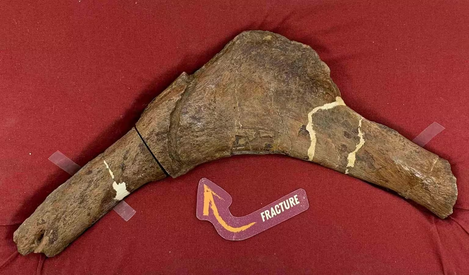
The vascular casts in this study were recovered from Scotty, the largest and one of the most complete T. rex specimens, curated at the Royal Saskatchewan Museum in Canada. The skeleton shows multiple injuries that likely accumulated over Scotty’s life about 66 million years ago. One rib bears a large, partially healed fracture; the blood-vessel network imaged there appears to record the increased vascular activity associated with bone healing.
Methods: synchrotron imaging and 3D reconstruction
Analyzing internal microstructures in fossil bone presents two main challenges: imaging the interior without destructive sampling, and penetrating very dense, mineral-replaced material. Medical CT scanners often lack the resolution and penetrating power needed for heavily mineralized fossils.
Synchrotron X-ray microtomography
The research team used synchrotron light—intense, highly collimated X-rays generated at particle accelerator facilities—to scan the rib at micron-scale resolution. Synchrotron microtomography can resolve fine three-dimensional details and also provide element-sensitive contrast, enabling chemical mapping of mineral phases.
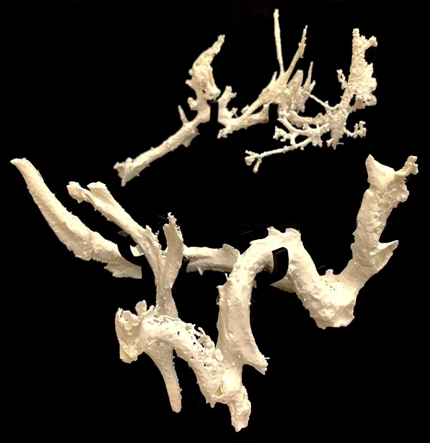
Chemical mapping showed the vascular canals preserved as iron-rich mineral casts, arranged in two distinct mineral layers. This layering reflects a complex diagenetic history—the sequence of chemical and environmental changes after burial—that led to exceptional preservation of microstructures.
Key discoveries and implications
The pattern of mineralized vessels corresponds with what is expected during active bone healing: a localized surge in vascularization to deliver cells and minerals to the repair site. By documenting these preserved angiogenic (vessel-forming) patterns, scientists gain direct anatomical evidence of how large theropods like T. rex responded to trauma.
Comparative analysis of vascular morphology could inform evolutionary questions: do vessel networks in dinosaurs resemble those of modern archosaurs (birds and crocodilians)? Can differences in vascular architecture be linked to growth rates, metabolism or immune response? This discovery also suggests a practical guide for future fieldwork—bones exhibiting healed injuries may be promising targets for locating additional soft-tissue traces.
Related technologies and future prospects
Applying particle-accelerator-based imaging, coupled with 3D modelling and non-destructive chemical analysis, is transforming paleontology. These cross-disciplinary methods allow researchers to interrogate fossils at cellular and microstructural scales while preserving specimens for future study. Data from mineralized soft tissues can be integrated with histology, isotopic analysis and comparisons to living relatives to reconstruct physiology and life history.
Expert Insight
"Finding preserved vascular casts in a T. rex rib gives us a direct window into healing processes in ancient animals," says Dr. Ana Ruiz, a paleobiologist not affiliated with the study. "It ties paleopathology to real physiological mechanisms, and helps us ask measurable questions about growth, immune response and behavior in extinct species."
Conclusion
The visualization of iron-rich blood-vessel casts in Scotty’s rib demonstrates how advanced imaging and chemical mapping can recover delicate biological signals from deep time. These findings expand our ability to infer dinosaur biology beyond bone shape alone—offering new data on healing, physiology and evolutionary relationships—and point toward targeted strategies for discovering further soft-tissue evidence in the fossil record.
Source: scitechdaily
Leave a Comment