4 Minutes
A tiny drama unfolded beneath the lens: immune cells edging closer, then slowly swallowing live melanoma cells whole. A single sequence of frames, and a long-held assumption about how our bodies police cancer shifted.
Researchers at the Garvan Institute of Medical Research in Sydney used intravital two-photon microscopy to watch a specific subgroup of macrophages—those that express the protein CD169—surround and actively engulf living melanoma cells in mice. This is not the slow cleanup crew clearing debris after the fact. These cells are attacking live tumor cells, nibbling away at the cancer along the tumor margin.
This is the first time macrophages have been recorded consuming live cancer cells in real time.
Macrophages commonly make up a large fraction of cells inside melanoma tumors, but until now their role has been murky. Some populations seem to shelter tumors; others may suppress them. By selectively removing CD169-positive macrophages, the team observed faster tumor growth, which suggested this subset plays a protective role at the tumor edge.
.avif)
Microscopy of a cross-section of mouse skin containing melanoma tumors. CD169+ macrophages are shown in green and yellow, forming a distinct boundary as they attempt to locally contain the cancer.
Why does that matter? Because these CD169 macrophages appear to act independently of the usual suspects—T cells and B cells—that most immunotherapies rely on. They form a physical and functional barrier, containing cancer cells and possibly flagging them to additional immune forces.
The finding gained extra weight after collaborators at the Melanoma Institute Australia examined human tissue and found the same CD169-positive cells hugging the borders of human melanomas and present in healthy skin. In short: what the microscope revealed in mice seems to have a counterpart in humans.
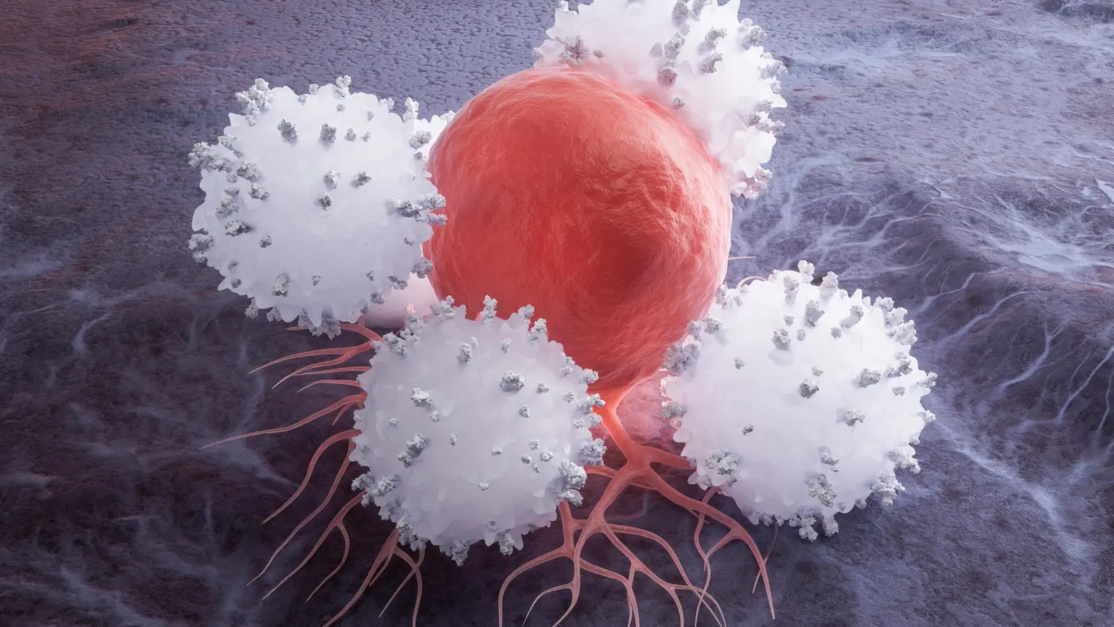
.avif)
A highly magnified view of a human melanoma tumor. Here, you can see the protective ‘housekeeping’ immune cells called CD169+ macrophages (highlighted in bright green and red) positioned right up against the cancer cells (in pink), ready to attack and engulf them.
There are clear clinical implications. Immune checkpoint inhibitors have transformed melanoma care, but many patients have 'cold' tumors that keep T cells out. Could these macrophages be coaxed into calling in reinforcements? The researchers suggest that after consuming tumor material, macrophages display fragments on their surface like a biological red flag, potentially recruiting T cells to finish the job.
Next steps are practical and ambitious. Scientists want to map how CD169 macrophages communicate with T cells, then figure out how to boost or reprogram them—either by increasing their numbers, enhancing their capacity to engulf cancer, or improving how they present tumor bits to T cells. Imagine treatments that mobilize an already-present immune contingent rather than importing a new army.
The discovery reorients how we view the tumor microenvironment: not just a battlefield between cancer and T cells, but a neighborhood where resident 'housekeepers' may quietly restrain malignancy. If these unsung defenders can be tuned, immunotherapy could be made effective for many more patients—and the strategy might extend beyond melanoma to other solid tumors rich in macrophages.
It began as a fleeting clip of cells moving under glass. Now the question is whether that footage can be turned into drugs that rally the immune system's own frontline.
Source: scitechdaily
Leave a Comment