
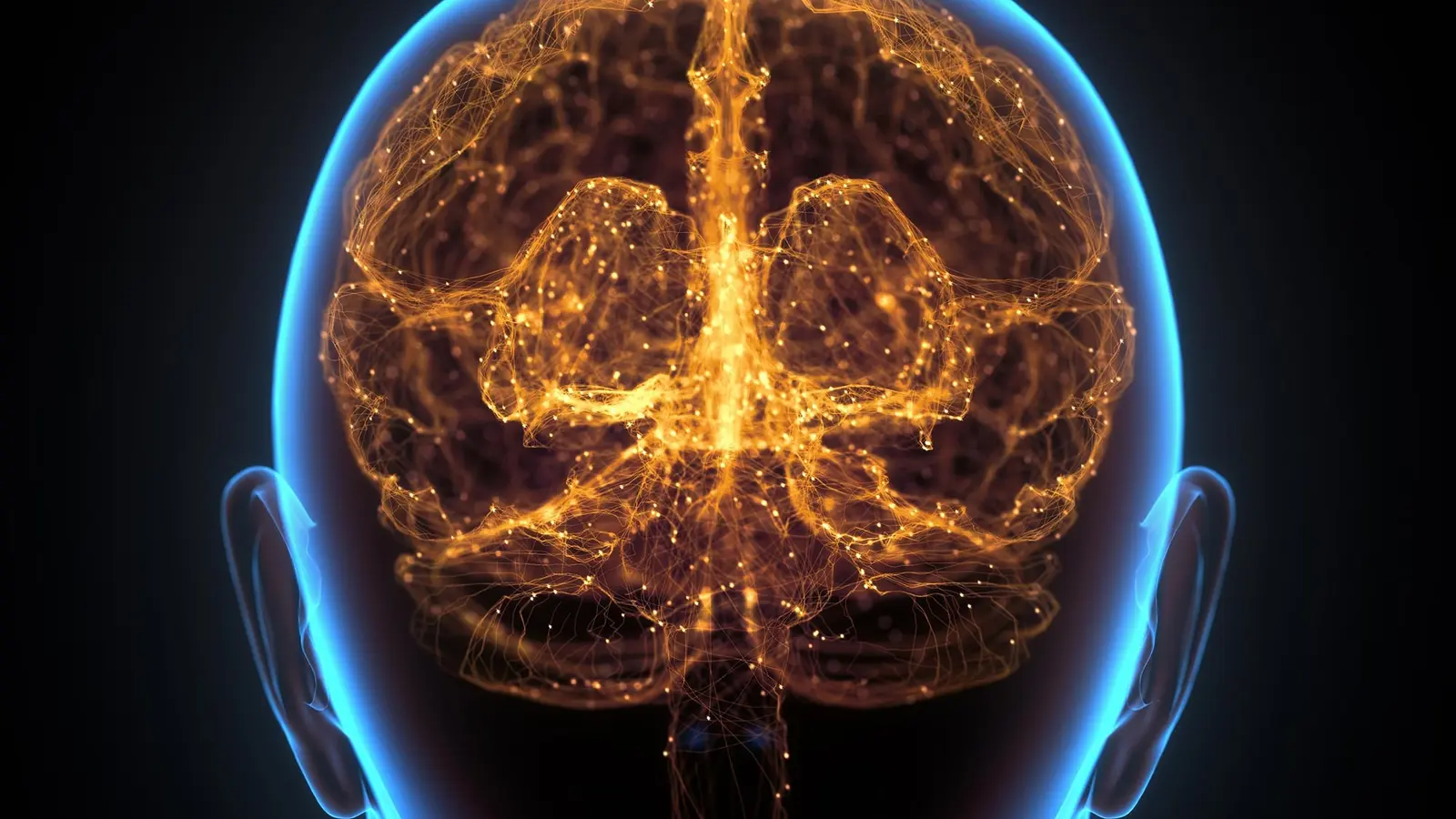
Imagine the skull as more than a protective shell. Picture it as a front-line outpost where immune cells gather, train, and launch swift counterattacks. That is the startling image emerging from a new study led by researchers at Washington University School of Medicine in St. Louis, published in Nature on August 19, 2026.
For decades neuroscientists treated the brain as insulated from routine immune activity. The new work upends that idea by identifying lymph node-like hubs inside the skull bone marrow of mice—microenvironments that react to brain tumors faster than distant lymph nodes do. The team also found traces of comparable immune cells in human skull marrow, suggesting the phenomenon may cross species.
What makes these hubs unusual is their location. Tiny channels connect brain tissue, the dura mater, and the skull marrow, allowing proteins, waste, and immune signals to move directly between the brain and the nearby marrow. When the researchers tracked molecules moving out of the brain, they observed them arriving in these skull niches, where specialized immune actors—organizing cells that resemble those in lymph nodes—are poised to respond.
Lead author Jang Hyun Park and senior author Jonathan Kipnis argue that the skull marrow is not passive. Park, a postdoctoral fellow at the Kipnis lab, said the team had never seen such complex immune structures in healthy bone marrow before. Kipnis, a distinguished professor at WashU Medicine, calls the discovery a rethink of how the nervous and immune systems talk to each other.
Researchers at WashU Medicine discovered lymph node-like structures (cyan) in the skull bone marrow of mice that play a role in mounting a rapid immune response in the brain.
Why does proximity matter? The researchers tested that directly in a glioblastoma model. When the skull immune hubs were disrupted pharmacologically, tumors advanced more rapidly and mice lived for shorter periods. Conversely, stimulating the skull marrow produced a much more vigorous local antibody response and slowed tumor growth. In an elegant experiment, a gel loaded with three immune-boosting proteins was placed beneath the scalp; immune activation flared first inside the skull hubs and only later in peripheral lymph nodes. Treated mice mounted stronger defenses and survived longer than controls.
The mechanisms resemble familiar immunology. Within those skull niches, cells akin to T follicular helper and B cells interact to generate high-output antibody production, the sort of coordinated response usually associated with lymph nodes. Except this time it happens right next to the organ those antibodies are meant to protect.
There are implications beyond brain cancer. If the skull houses rapid-response immune stations, could diseases with an immune component—Alzheimer's, Parkinson's, long COVID, or certain psychiatric conditions—be influenced directly by activity in these marrow hubs? Kipnis suggests therapeutic strategies could one day target the skull spaces themselves, delivering modulators locally and avoiding widespread peripheral side effects.
Of course, mice are not humans. The study reports human skull marrow contains related immune cells, but much remains to be mapped: the precise cell types, how these hubs form, and whether therapies can safely and reliably access them in people. Still, the notion that the brain has neighbors acting as first responders changes how we frame neuroimmunology.
Curious? Good. This discovery opens as many questions as it answers, and researchers are already sketching the next experiments to see how deep the skulls secrets run.


Leave a Comment
Comments
No comments yet. Be the first.