4 Minutes
Light touch. A brush of fabric. For millions living with neuropathic pain, those ordinary sensations can become electric jolts of suffering. What if the pain is less about nerves misfiring and more about a slow-burning energy failure inside the cells themselves?
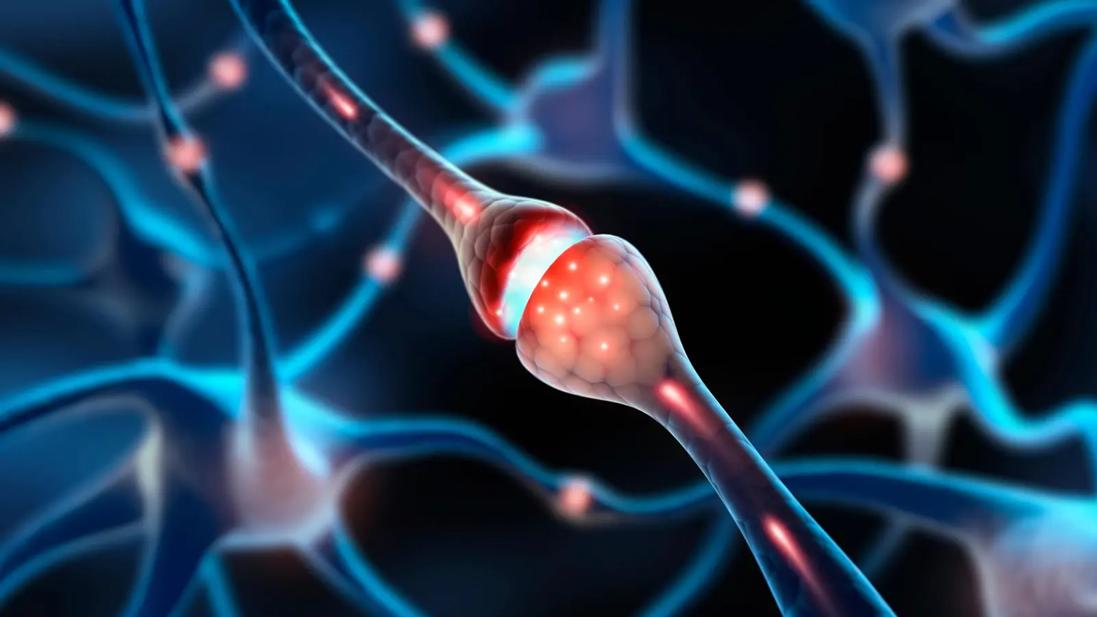
Chronic nerve pain may stem from a hidden energy crisis inside damaged cells. In a new study, researchers explore whether restoring the tiny power generators within nerves can reverse pain rather than just dull it. Credit: Shutterstock
How broken batteries make nerves hurt
Mitochondria are tiny but critical: they produce the ATP that cells use to power ion pumps, repair enzymes, and other housekeeping tasks. When those powerhouses falter, neurons lose their ability to maintain membrane potential and to fight inflammation. The result can be persistent, abnormal pain signaling — the hallmark of neuropathic conditions such as diabetic neuropathy and chemotherapy-induced neuropathy.
Researchers at Duke University School of Medicine tested a bold idea: rather than only suppressing pain signals with drugs, why not restore healthy mitochondria to wounded nerve cells and address the energetic root cause? The team used human tissue and mouse models to probe that question, and the results are striking.
In live experiments, boosting mitochondrial function — either by encouraging intercellular transfer or by directly supplying healthy mitochondria — lowered pain behaviors substantially. In some tests, relief lasted up to 48 hours. In others, behavioral signs of pain fell by roughly half. Those are not mere numbers; they point to a mechanistic shift in how we might treat chronic neuropathic pain.
Cells lending a hand: satellite glia and nanotubes
The study highlights an underappreciated form of cellular cooperation. Sensory neurons are surrounded by satellite glial cells. Once considered passive support, these glia appear to pass mitochondria to neurons through slender conduits called tunneling nanotubes. Think of them as microscopic bridges, ferrying energy packets from one cell to another.
When the transfer breaks down, distal nerve endings — especially in hands and feet — begin to decay. Tingling, numbness and hypersensitivity follow. In mouse models, enhancing the mitochondrial handoff reduced pain behaviors significantly. In a more direct experiment, investigators injected isolated mitochondria into the dorsal root ganglia, the clusters of nerve cell bodies that shuttle sensory signals to the brain. When the grafted mitochondria were healthy, pain measures improved. When mitochondria came from diabetic donors, the effect vanished, underscoring that quality matters.
At the molecular level, the protein MYO10 emerged as essential for building tunneling nanotubes. Without the scaffolding and motor proteins that shape these bridges, the energy exchange falters. The discovery frames a new target: if scientists can boost MYO10-driven nanotube formation or otherwise facilitate mitochondrial transfer, they might restore neuronal vigor without systemic side effects.
Pain researcher Ru-Rong Ji, PhD, director of the Center for Translational Pain Medicine at Duke, frames the approach like this: "Give damaged nerves fresh mitochondria — or help them make more of their own — and you can reduce inflammation and support healing." It is a different logic from merely damping pain signals. It is restorative.
What this means and what remains unknown
The implications are broad. If mitochondrial transfer is a general support mechanism, it could influence obesity, stroke recovery, cancer biology and multiple degenerative conditions. For neuropathic pain specifically, therapies that coax glial cells to donate mitochondria, stabilize transferred organelles, or deliver healthy mitochondria directly could become complementary options to drugs and neuromodulation.
But caution is warranted. The experiments relied on animal models and ex vivo human tissue. High-resolution imaging of living nerve tissue will be needed to map how nanotubes form, how mitochondria travel, and how long donated organelles remain functional inside recipient neurons. Safety questions also arise: how to target mitochondria where they are needed without provoking immune responses or off-target effects. And the variable quality of donor mitochondria — as seen with diabetic samples — means personalized approaches may be required.
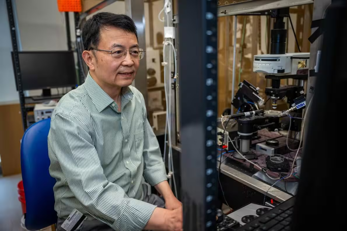
Pain researcher Ru-Rong Ji, PhD, in his anesthesiology lab at the Duke University School Medicince.
Expert Insight
Dr. Maya El-Amin, a cellular neurobiologist at a major research hospital (commenting as an independent expert): "This work reframes neuropathic pain as a metabolic problem as much as a neural one. The idea that glial cells can donate mitochondria implies an active support network we underestimated. Translating that into treatments will take time, but the roadmap is clearer now — focus on organelle quality, delivery routes, and the cellular machinery that builds those nanotubes."
Mitochondria, once relegated to textbooks as static powerhouses, are proving to be dynamic players in tissue health. Fix their flow, and you may fix pain where it starts — deep inside the cell.
Comments
RaMox
Quick q: injecting mitochondria into DRG - is immune rejection a big deal? sounds promising but seems risky, targeting seems hard. anyone know?
cellXen
wow this flips everything i thought about neuropathic pain. Mitochondrial transfer? mind blown, hopeful but nervous. Curious how durable the fixes are, if they really last.
Leave a Comment